. 2024 Feb 16;134(4):393-410.
doi: 10.1161/CIRCRESAHA.123.322729. Epub 2024 Jan 26.

Protein Kinase A Is a Master Regulator of Physiological and Pathological Cardiac Hypertrophy

Affiliations

Protein Kinase A Is a Master Regulator of Physiological and Pathological Cardiac Hypertrophy

Yingyu Bai et al. Circ Res. .

Abstract

Background: The sympathoadrenergic system and its major effector PKA (protein kinase A) are activated to maintain cardiac output coping with physiological or pathological stressors. If and how PKA plays a role in physiological cardiac hypertrophy (PhCH) and pathological CH (PaCH) are not clear.

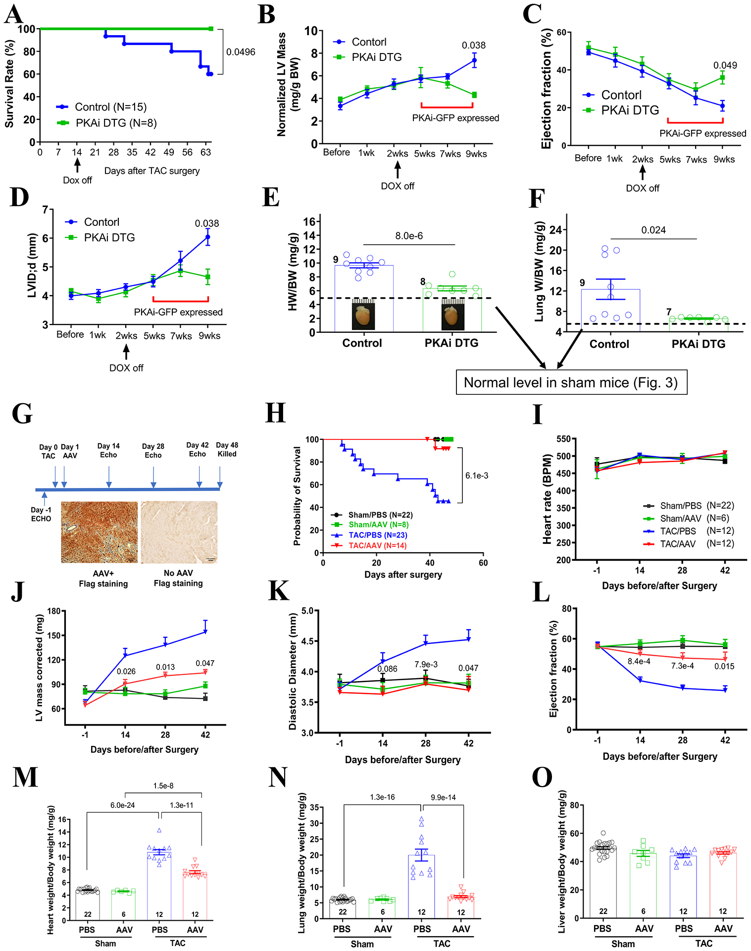
Methods: Transgenic mouse models expressing the PKA inhibition domain (PKAi) of PKA inhibition peptide alpha (PKIalpha)-green fluorescence protein (GFP) fusion protein (PKAi-GFP) in a cardiac-specific and inducible manner (cPKAi) were used to determine the roles of PKA in physiological CH during postnatal growth or induced by swimming, and in PaCH induced by transaortic constriction (TAC) or augmented Ca2+ influx. Kinase profiling was used to determine cPKAi specificity. Echocardiography was used to determine cardiac morphology and function. Western blotting and immunostaining were used to measure protein abundance and phosphorylation. Protein synthesis was assessed by puromycin incorporation and protein degradation by measuring protein ubiquitination and proteasome activity. Neonatal rat cardiomyocytes (NRCMs) infected with AdGFP (GFP adenovirus) or AdPKAi-GFP (PKAi-GFP adenovirus) were used to determine the effects and mechanisms of cPKAi on myocyte hypertrophy. rAAV9.PKAi-GFP was used to treat TAC mice.

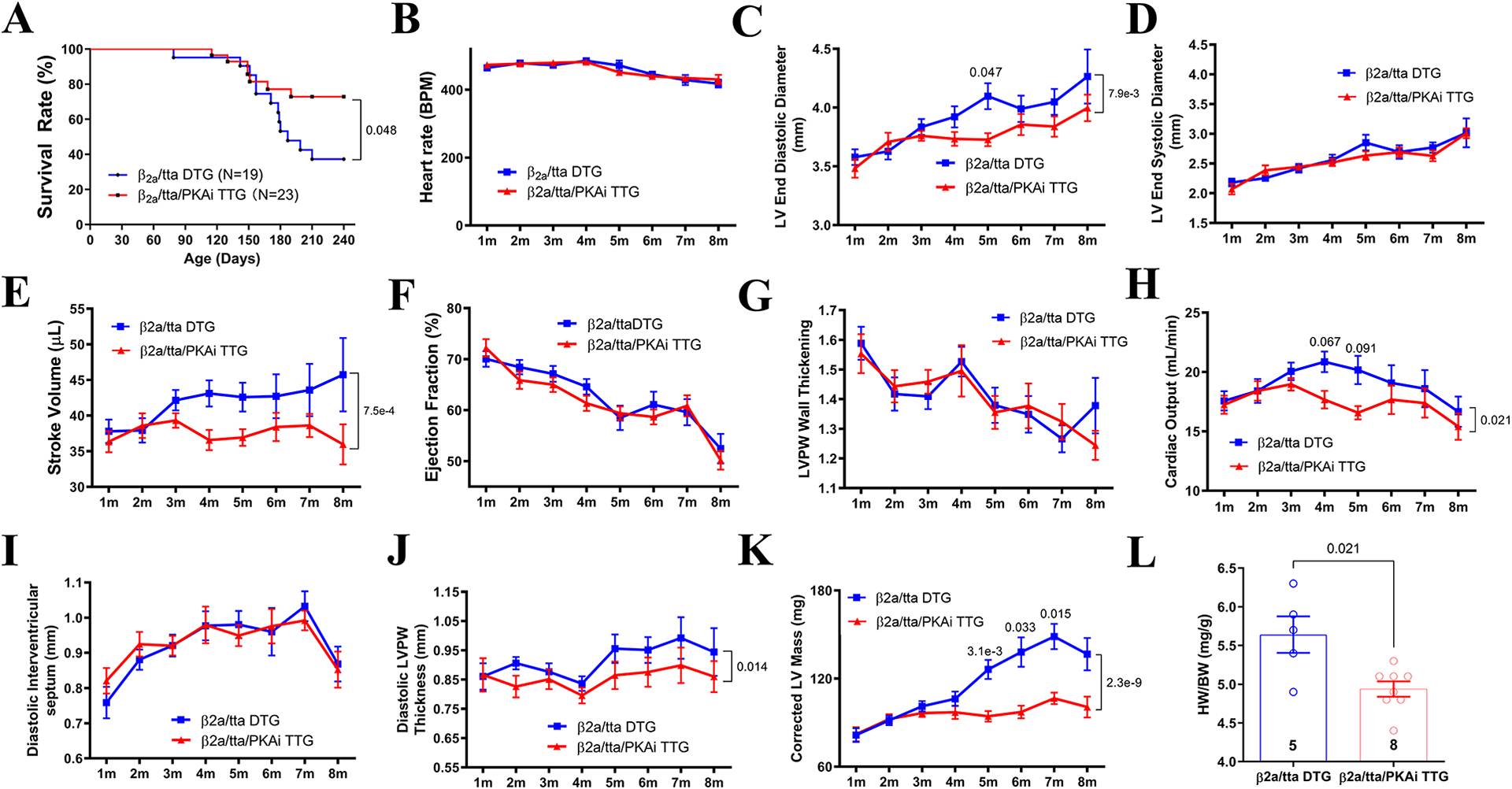
Results: (1) cPKAi delayed postnatal cardiac growth and blunted exercise-induced PhCH; (2) PKA was activated in hearts after TAC due to activated sympathoadrenergic system, the loss of endogenous PKIα (PKA inhibition peptide α), and the stimulation by noncanonical PKA activators; (3) cPKAi ameliorated PaCH induced by TAC and increased Ca2+ influxes and blunted neonatal rat cardiomyocyte hypertrophy by isoproterenol and phenylephrine; (4) cPKAi prevented TAC-induced protein synthesis by inhibiting mTOR (mammalian target of rapamycin) signaling through reducing Akt (protein kinase B) activity, but enhancing inhibitory GSK-3α (glycogen synthase kinase-3α) and GSK-3β signals; (5) cPKAi reduced protein degradation by the ubiquitin-proteasome system via decreasing RPN6 phosphorylation; (6) cPKAi increased the expression of antihypertrophic atrial natriuretic peptide (ANP); (7) cPKAi ameliorated established PaCH and improved animal survival.

Conclusions: Cardiomyocyte PKA is a master regulator of PhCH and PaCH through regulating protein synthesis and degradation. cPKAi can be a novel approach to treat PaCH.

Keywords: cyclic AMP-dependent protein kinases; heart; hypertrophy; mechanistic target of rapamycin complex 1; proteolysis.


Conflict of interest statement

Disclosures None.

Figures

Figure 1.
Figure 1.
Cardiac PKAi-GFP (protein kinase A inhibitor α amino acids 1–25 fused with the green fluorescent protein) reduces ventricular myocyte developmental growth and blunts physiological cardiac hypertrophy induced by exercise. A. An immunostaining image of GFP expression in a VLE mouse heart tissue showing mosaic expression of GFP. B. Images of green (PKAi-GFP+) and nongreen (PKAi-GFP) cardiomyocytes isolated from the same heart taken under bright light and GFP-excitation light. Myocytes were isolated from 4-month-old control and DTG mice with mosaic PKAi-GFP expression. C. Surface area of green and non-green myocytes isolated from the same hearts. D. An immunostaining image of GFP expression in a HE mouse heart tissue section showing high expression of GFP homogenously in all ventricular myocytes. E. The HW/BW ratio of 4-month-old control and PKAi-GFP high expression DTG mice. F-G. Surface areas of isolated ventricular myocytes of control and PKAi-GFP HE DTG mice. H. Control and PKAi-GFP ME DTG mice were subjected to swimming, 2 hours in the morning and 2 hours in the afternoon (totally 4 hours/day) for 3 weeks. I-L. Body weights and thickness of interventricular septum (IVS; d), left ventricular posterior (free) wall (LVPW; d) during diastole, corrected LV mass measured with echocardiography, showing increased thickness of these walls in control mice but not in TG mice subjected to exercise. M. HW/BW ratios of hearts of control and PKAi-GFP medium expression mice, showing cPKAi blunted exercise induced cardiac hypertrophy. P values were reported above the comparison lines. Animal numbers were added in the bars. Student’s t test was used in C; Mann-Whitney test was used for E and G; two-way ANOVA with post-hoc test (Bonferroni correction) was used in I through M. The scale bars in all images were 100 μm.
Figure 2.
Figure 2.
Increased PKA activity in the heart after pressure overload (TAC) due to SAS activation, noncanonical activator (ROS and AngII) stimulation, and the loss of endogenous PKIα. A. HW/BW ratio at different time points after TAC. B. Basal PKA activity in cardiac tissue homogenates of sham or TAC mice at different times after surgeries. C. Maximal PKA activity activated by 1 μM exogenous cAMP in cardiac tissue homogenates as in B. D. PKA activation by H2O2 and angiotensin II (AngII) was blocked by cPKAi. E. Immunostaining of PKIα in sham and TAC cardiac tissue sections. P values were reported above the comparison lines. Animal numbers were added in or next to the bars. Kruskal-Wallis test was used for A; aligned rank transform followed by two-way ANOVA with post-hoc (Bonferroni correction) was used for B-D. Scale bars in E were 25 μm.
Figure 3.
Figure 3.. Medium level of PKAi-GFP expression blunts PaCH and improves cardiac function after TAC.
A. Pressure gradients across the transverse aortic constriction were similar in control TAC mice (60.5±2.5mmHg) and in PKAi-GFP/ttA DTG TAC mice (62.3±3.1mmHg). B. PKAi-GFP DTG mice survived better after TAC. When echocardiography (ECHO) was performed, the heart rates were controlled to be similar (C). TAC significantly decreased ejection fraction (D) and increased LV diastolic diameter (E) in control mice but not in DTG mice. F-I. TAC significantly increased end diastolic volume (F) and end systolic volume (G) in control mice but not in DTG mice, thus leading to a decreased stroke volume (H) and a worse cardiac output (I). J-L. At 8 weeks post TAC, DTG mice had reduced cardiac hypertrophy (corrected LV mass (J), HW/BW ratio (K) and lung weight to body weight ratio (L), an index of cardiac decompensation. M. Contractions of myocytes isolated from control and DTG hearts subjected to sham and TAC surgeries for 8 weeks. Ma, Representative cardiomyocyte contraction traces. Mb, Fractional shortening amplitudes. Mc, Time to peak. Md, Time to half relaxation. Me, maximal contraction/relaxation rate. N. Ca2+ transients of myocytes isolated from control and DTG hearts subjected to sham and TAC surgeries for 8 weeks. Na, Representative cardiomyocyte Ca2+ transients traces. Nb, Diastolic Ca2+. Nc, Systolic Ca2+. Nd, Ca2+ transient amplitudes. Ne, Time to peak. Nf, Time to half relaxation. Ng, Ca2+ transient decay time constant (Tau). O & P. Myocyte cross sectional area of papillary muscles of control and TG hearts after TAC. Q & R. Masson’s Trichrome staining of cardiac tissue of sham or TAC operated control and PKAi-GFP DTG animals. TAC significantly induced cardiac fibrosis in control mice but not in PKAi-GFP DTG mice. P values were reported above the comparison lines. Animal numbers were added in or next to the bars for A-L, P and R. The numbers next to the bars in M and N panels were myocyte numbers from 3 or 4 animals depicted in M.a. Student’s t test was used for A; the difference in survival rates was determined by Kaplan-Meier survival analysis (B); aligned rank transform followed by two-way ANOVA with post-hoc test (Bonferroni correction) was used for C-L, P and R; Nested ANOVA analyses were done for M and N pannels .
Figure 4.
Figure 4.. cPKAi reduces PaCH induced by increased Ca2+ influx.
PKAi-GFP ME TG mice were crossbred with Cavβ2a/ttA DTG mice. A. Triple TG (TTG) mice survived better than Cavβ2a/ttA DTG mice. Heart rates were controlled similar between groups for ECHO (B). cPKAi significantly reduced the dilation of the LV chamber (C), without affecting LV end systolic diameter (D). cPKAi significantly blunted the dilation, leading to a decreased stroke volume (E) and cardiac output (H) without affecting LVPW wall thickening (G) and ejection fraction (F). cPKAi did not affect diastolic septum thickness (I) but blunted LVPW thickening induced by increased ICa-L (J), resulting into a reduced corrected LV mass (K). Gravimetric analysis showed a reduced HW/BW (L) by cPKAi in TTG animals euthanized at 8m of age. P values were reported above the comparison lines. Animal numbers were added in or next to the bars; animal numbers used in A-L varied as animals died during experiments. Data from these animals were not excluded. The difference in survival rates was determined by Kaplan-Meier survival analysis; two-way ANOVA with post-hoc test (Bonferroni correction) was used in C through K; Mann-Whitney test was used in L.
Figure 5.
Figure 5.. cPKAi blunts protein synthesis induced by TAC.
A & B. De novo protein synthesis was quantitated by the incorporation of puromycin into nascent proteins (darker lanes); S: sham; T: TAC. C & D. Total and phosphorylation of key regulatory molecules involved in protein synthesis in control hearts and in PKAi-GFP DTG hearts subjected to pressure overload (TAC) or sham surgery. P values were reported above the comparison lines. Animal numbers were added in or next to the bars. Aligned rank transform followed by two-way ANOVA with post-hoc test (Bonferroni correction) was used in B; two-way ANOVA with post-hoc test (Bonferroni correction) was used in other comparisons in this figure.
Figure 6.
Figure 6.. Protein degradation is reduced in cPKAi hearts after TAC.
A & B. Total ubiquitination (A) and poly ubiquitination (B) of proteins was increased by TAC in control mice, which was blunted by cPKAi. C. 20S proteasome activities were increased by TAC in control mice but not in cPKAi DTG mice. D. The phosphorylation of Rpn6 by PKA, a key mediator of PKA-regulated proteasome activity, was increased by TAC in control mice but not in PKAi-GFP DTG mice. P values were reported above the comparison lines. Animal numbers were added in or next to the bars. Aligned rank transform followed by two-way ANOVA with post-hoc test (Bonferroni correction) was used in C.
Figure 7.
Figure 7.. cPKAi inhibits cardiomyocyte hypertrophy partially through increasing antihypertrophic ANP expression.
Neonatal rat cardiomyocytes (NRCMs) were infected with AdGFP or AdPKAi-GFP and then treated with isoproterenol (ISO), phenylephrine (PE) or angiotensin II (AngII). NCRM hypertrophy induced by ISO, PE and AngII as indicated by the increases in myocyte surface area (A) and protein synthesis (protein/DNA ratio, B) was prevented by PKAi-GFP. Without hypertrophic stimulation, cPKAi increased ANP mRNA expression in NRCMs (C), pro-ANP expression in hearts of PKAi-GFP DTG mice (D) and ANP concentration in serum of PKAi-GFP DTG mice (E). F&G. The PKAi-GFP mediated antihypertrophic effect was blunted by ANP neutralizing antibody. F. The representative images of GFP positive NRCMs treated with or without ANP neutralizing antibody when challenge with hypertrophic stimuli (F). Cell images were taken with adjusted exposure time to make the image fluorescence brightness to the same to facilitate surface area measurements. The quantitation of cell surface area of NRCMs (G). P value was shown above each bracket. Aligned rank transform followed by two-way ANOVA with post-hoc test (Bonferroni correction) was used in A-C and G;Mann-Whitney test was used in D and E. The scale bars in F are 25μm. The numbers next to datasets in D & E are animal numbers used and in A, B, C, G are independent experiment times performed.
Figure 8.
Figure 8.. cPKAi reverses PaCH induced by TAC.
A-F. In the PKAi-GFP HE DTG mice, the expression of PKAi-GFP was kept off by feeding the mice on Dox-containing water and PaCH was induced by TAC for 2 weeks. Then PKAi-GFP expression was induced by removing the Dox-containing water. cPKAi at 5 weeks after TAC prevented animal death (A), partially reversed cardiac hypertrophy (B, LV mass determined by ECHO; E, HW/BW), and halted cardiac functional decompensation (C, D and F). G-O. AAV mediated cPKAi gene therapy to treat PaCH. G. The scheme of the gene therapy approach and PKAi-GFP expression in the heart showing almost all myocytes were expressing PKAi-GFP-6x FLAG. Animal survival was significantly improved with a single dose of AAV (H). Heart rates were similar for ECHO (I). rAAV9.PKAi-GFP treatment reduced corrected LV mass (J) and the dilation of the LV chamber (K) and improved ejection fraction after TAC surgeries (L). Gravimetric analysis showed there was a reduced HW/BW (M) and a reduced LungW/BW (N) in the rAAV9.PKAi-GFP-TAC group. There was nothing different of LiverW/BW (O) among all the groups. P value was shown above each bracket. Survival rate difference was determined by Kaplan-Meier survival analysis in A and H; two-way ANOVA with post-hoc test (Bonferroni correction) was used in B through D and I through O;Student’s t test was used in E&F. The scale bars in F are 100μm. The numbers next to datasets are animal numbers used.

References

    1. Nakamura M, Sadoshima J. Mechanisms of physiological and pathological cardiac hypertrophy. Nat Rev Cardiol. 2018;15:387–407 - PubMed
    1. Liu Y, Chen J, Fontes SK, Bautista EN, Cheng Z. Physiological and pathological roles of protein kinase a in the heart. Cardiovasc Res. 2022;118:386–398 - PMC - PubMed
    1. Frey N, Katus HA, Olson EN, Hill JA. Hypertrophy of the heart: A new therapeutic target? Circulation. 2004;109:1580–1589 - PubMed
    1. Wang J, Gareri C, Rockman HA. G-protein-coupled receptors in heart disease. Circ Res. 2018;123:716–735 - PMC - PubMed
    1. Antos CL, Frey N, Marx SO, Reiken S, Gaburjakova M, Richardson JA, Marks AR, Olson EN. Dilated cardiomyopathy and sudden death resulting from constitutive activation of protein kinase a. Circ Res. 2001;89:997–1004 - PubMed

Publication types

Substances