. 2023 Dec 21;14(1):14.
doi: 10.3390/biom14010014.

Evaluation of Myocilin Variant Protein Structures Modeled by AlphaFold2

Affiliations

Evaluation of Myocilin Variant Protein Structures Modeled by AlphaFold2

Tsz Kin Ng et al. Biomolecules. .

Abstract

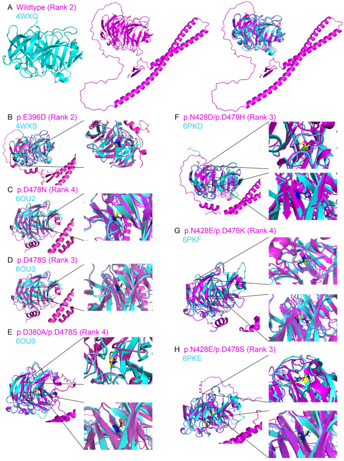
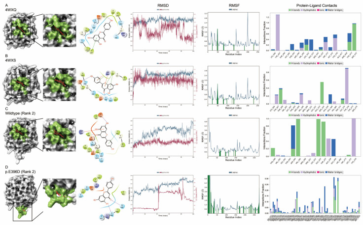
Deep neural network-based programs can be applied to protein structure modeling by inputting amino acid sequences. Here, we aimed to evaluate the AlphaFold2-modeled myocilin wild-type and variant protein structures and compare to the experimentally determined protein structures. Molecular dynamic and ligand binding properties of the experimentally determined and AlphaFold2-modeled protein structures were also analyzed. AlphaFold2-modeled myocilin variant protein structures showed high similarities in overall structure to the experimentally determined mutant protein structures, but the orientations and geometries of amino acid side chains were slightly different. The olfactomedin-like domain of the modeled missense variant protein structures showed fewer folding changes than the nonsense variant when compared to the predicted wild-type protein structure. Differences were also observed in molecular dynamics and ligand binding sites between the AlphaFold2-modeled and experimentally determined structures as well as between the wild-type and variant structures. In summary, the folding of the AlphaFold2-modeled MYOC variant protein structures could be similar to that determined by the experiments but with differences in amino acid side chain orientations and geometries. Careful comparisons with experimentally determined structures are needed before the applications of the in silico modeled variant protein structures.

Keywords: AlphaFold2; molecular simulation; myocilin; protein structure; variants.


Conflict of interest statement

The authors declare no potential conflicts of interest.

Figures

Figure 1
Figure 1
Structure similarity analysis on the AlphaFold2-predicted variant protein structures to the experimentally determined structures from the protein data bank. The 5 AlphaFold2-predicted protein structures (Rank 0–Rank 4) of variants from the protein data bank, (A) 6SSO, (B) 7K77, and (C) 7RLG chain A, were aligned with the corresponding experimentally determined protein structures from the protein data bank (PDB). The alignment of the AlphaFold2-predicted variant protein structures with the highest template modeling score is shown on the right. Green: the PDB structure; Cyan: the AlphaFold2 modeled Rank 0 structure; Magentas: the AlphaFold2 modeled Rank 1 structure; Yellow: the AlphaFold2 modeled Rank 2 structure; Salmon: the AlphaFold2 modeled Rank 3 structure; Light gray: the AlphaFold2 modeled Rank 0 structure; Red: the variant amino acid side chain of the PDB structure; Blue: the variant amino acid side chain of the AlphaFold2 modeled Rank 0 structure; Orange: the variant amino acid side chain of the AlphaFold2 modeled Rank 1 structure; Dark gray: the variant amino acid side chain of the AlphaFold2 modeled Rank 2 structure; Brown: the variant amino acid side chain of the AlphaFold2 modeled Rank 3 structure; Purple: the variant amino acid side chain of the AlphaFold2 modeled Rank 4 structure.
Figure 2
Figure 2
Structure similarity analysis on the AlphaFold2-predicted myocilin wildtype and variant protein structures to the experimentally determined structures. The AlphaFold2-predicted myocilin (A) wildtype and variant protein structures ((B) p.E396D; (C) p.D478N; (D) p.D478S; (E) p.D380A/p.D478S; (F) p.N428D/p.D478H; (G) p.N428E/p.D478K; (H) p.N428E/p.D478S) with highest template modeling score (red) were aligned with the corresponding experimentally determined protein structures from the protein data bank (green). Yellow: the side chain of the amino acid residue from the experimentally determined protein structure. Blue: the side chain of the amino acid residue from the AlphaFold2-predicted protein structure.
Figure 3
Figure 3
Structure similarity analysis of the experimentally determined myocilin wildtype and variant protein structures. The experimentally determined myocilin variant protein structures ((A) p.E396D; (B) p.D478N; (C) p.D478S; (D) p.D380A/p.D478S; (E) p.N428D/p.D478H; (F) p.N428E/p.D478K; (G) p.N428E/p.D478S) (red) were aligned with the experimentally determined myocilin wildtype protein structure (4WXQ) from the protein data bank (green). Yellow: the side chain of the amino acid residue from the wildtype protein structure. Blue: the side chain of the amino acid residue from the variant protein structures.
Figure 4
Figure 4
Structure similarity analysis of the AlphaFold2-predicted myocilin wildtype and variant C-terminus protein structures with the protein data bank identities. The AlphaFold2-predicted C-terminus protein structures of myocilin variants ((A) p.E396D; (B) p.D478N; (C) p.D478S; (D) p.D380A/p.D478S; (E) p.N428D/p.D478H; (F) p.N428E/p.D478K; (G) p.N428E/p.D478S) with the protein data bank identities (red) were aligned with the AlphaFold2-predicted myocilin wildtype protein structure (Rank 2) (green). Yellow: the side chain of the amino acid residue from the wild-type protein structure. Blue: the side chain of the amino acid residue from the variant protein structures.
Figure 5
Figure 5
Structure similarity analysis of the AlphaFold2-predicted C-terminus structures of myocilin wildtype and variant proteins without experimentally determined structures. The AlphaFold2-predicted C-terminus protein structures of myocilin variants ((A) p.C245Y; (B) p.G252R; (C) p.S313F; (D) p.E323K; (E) p.T353I; (F) p.G367R; (G) p.Q368*; (H) p.P370L; (I) p.D384H; (J) p.A488V) without experimentally determined structures (red) were aligned with the AlphaFold2-predicted myocilin wildtype protein structure (Rank 2) (green). Yellow: the side chain of the amino acid residue from the wildtype protein structure. Blue: the side chain of the amino acid residue from the variant protein structures. Brown: disulfide bond.
Figure 6
Figure 6
Molecular dynamics analysis on the experimentally determined and AlphaFold2-predicted myocilin wildtype and variant protein structures. (A,E,I,M) Root-mean-square deviation (RMSD) analysis. (B,F,J,N) Root-mean-square fluctuation (RMSF) analysis. (C,G,K,O) residue cross-correlation analysis. (D,H,L,P) normal mode analysis. (A,B) Comparison of the olfactomedin-like domain of the AlphaFold2-predicted wildtype myocilin protein structure (Rank 2) to that of the experimentally determined wild-type myocilin protein structure (4WXQ). (E,F) Comparison of the olfactomedin-like domain of the AlphaFold2-predicted myocilin p.E396D variant protein structure (Rank 2) to that of the experimentally determined myocilin p.E396D variant protein structure (4WXS). (I,J) Comparison of the olfactomedin-like domain of the experimentally determined myocilin p.E396D variant protein structure (4WXS) to that of the experimentally determined wild-type myocilin protein structure (4WXQ). (M,N) Comparison of the olfactomedin-like domain of the AlphaFold2-predicted myocilin p.E396D variant protein structure (Rank 2) to that of the AlphaFold2-predicted wild-type myocilin protein structure (Rank 2). (C,D) The experimentally determined wildtype myocilin protein structure (4WXQ). (G,H) The experimentally determined myocilin p.E396D variant protein structure (4WXS). (K,L) The AlphaFold2-predicted wildtype myocilin protein structure (Rank 2). (O,P) The AlphaFold2-predicted myocilin p.E396D variant protein structure (Rank 2).
Figure 7
Figure 7
Molecular docking analysis of apigenin on the experimentally determined and AlphaFold2-predicted myocilin wildtype and variant protein structures. Molecular docking analysis of apigenin (red) on the experimentally determined myocilin (A) wildtype (4WXQ) and (B) variant (4WXS; p.E396D) protein structures and the AlphaFold2-predicted C-terminus protein structures of myocilin (C) wild-type (Rank 2) and (D) p.E396D variant (Rank 2). The surface structural representation, binding site (green), protein–ligand root-mean-square deviation (RMSD), protein root-mean-square fluctuation (RMSF), and protein–ligand contacts are shown.
Figure 8
Figure 8
Molecular docking analysis of Gw5074 on the experimentally determined and AlphaFold2-predicted myocilin wildtype and variant protein structures. Molecular docking analysis of Gw5074 (orange) on the experimentally determined myocilin (A) wildtype (4WXQ) and (B) variant (4WXS; p.E396D) protein structures and the AlphaFold2-predicted C-terminus protein structures of myocilin (C) wild-type (Rank 2) and (D) p.E396D variant (Rank 2). The surface structural representation, binding site (green), protein–ligand root-mean-square deviation (RMSD), protein root-mean-square fluctuation (RMSF), and protein–ligand contacts are shown.

References

    1. Kapetanakis V.V., Chan M.P., Foster P.J., Cook D.G., Owen C.G., Rudnicka A.R. Global variations and time trends in the prevalence of primary open angle glaucoma (POAG): A systematic review and meta-analysis. Br. J. Ophthalmol. 2016;100:86–93. doi: 10.1136/bjophthalmol-2015-307223. - DOI - PMC - PubMed
    1. Weinreb R.N., Aung T., Medeiros F.A. The pathophysiology and treatment of glaucoma: A review. JAMA. 2014;311:1901–1911. doi: 10.1001/jama.2014.3192. - DOI - PMC - PubMed
    1. Kubota R., Noda S., Wang Y., Minoshima S., Asakawa S., Kudoh J., Mashima Y., Oguchi Y., Shimizu N. A novel myosin-like protein (myocilin) expressed in the connecting cilium of the photoreceptor: Molecular cloning, tissue expression, and chromosomal mapping. Genomics. 1997;41:360–369. doi: 10.1006/geno.1997.4682. - DOI - PubMed
    1. Stone E.M., Fingert J.H., Alward W.L., Nguyen T.D., Polansky J.R., Sunden S.L., Nishimura D., Clark A.F., Nystuen A., Nichols B.E., et al. Identification of a gene that causes primary open angle glaucoma. Science. 1997;275:668–670. doi: 10.1126/science.275.5300.668. - DOI - PubMed
    1. Gupta V., Somarajan B.I., Gupta S., Walia G.K., Singh A., Sofi R., Chaudhary R.S., Sharma A. The mutational spectrum of Myocilin gene among familial versus sporadic cases of Juvenile onset open angle glaucoma. Eye. 2021;35:400–408. doi: 10.1038/s41433-020-0850-z. - DOI - PMC - PubMed

Publication types

LinkOut - more resources