. 2024 Jan 19;12(1):203.
doi: 10.3390/microorganisms12010203.

A Three-Dimensional Model of Bacterial Biofilms and Its Use in Antimicrobial Susceptibility Testing

Affiliations

A Three-Dimensional Model of Bacterial Biofilms and Its Use in Antimicrobial Susceptibility Testing

Hala R Ali et al. Microorganisms. .

Abstract

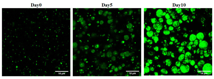
(1) Background: The discrepant antimicrobial susceptibility between planktonic and biofilm bacterial modes poses a problem for clinical microbiology laboratories and necessitates a relevant 3D experimental model allowing bacteria to grow in biofilm mode, in vitro, for use in anti-biofilm susceptibility testing. (2) Methods: This work develops a 3D biofilm model consisting of alginate beads containing S. aureus biofilm and encased within two thick layers of alginate matrix. The constructed model was placed on a thin Boyden chamber insert suspended on a 24-well culture plate containing the culture medium. The antibacterial activity of bacitracin and chlorhexidine digluconate (CD), either combined or separately, against 2D S. aureus culture was compared to that in the 3D biofilm model. Quantitative analysis and imaging analysis were performed by assessing the bacterial load within the matrix as well as measuring the optical density of the culture medium nourishing the matrix. (3) Results: The 3D biofilm model represented the typical complex characteristics of biofilm with greater insusceptibility to the tested antimicrobials than the 2D culture. Only bacitracin and CD in combination at 100× the concentration found to be successful against 2D culture were able to completely eliminate the 3D biofilm matrix. (4) Conclusions: The 3D biofilm model, designed to be more clinically relevant, exhibits higher antimicrobial insusceptibility than the 2D culture, demonstrating that the model might be useful for testing and discovering new antimicrobial therapies. The data also support the view that combination therapy might be the optimal approach to combat biofilm infections.

Keywords: 3D biofilm model; S. aureus biofilm; antimicrobial susceptibility testing; chronic wound infection; combination therapy.


Conflict of interest statement

The authors declare no conflicts of interest.

Figures

Figure 1
Figure 1
Construction of biofilm model: (a) S. aureus biofilm in 96-well plates; (b) S. aureus alginate beads; (c) biofilm beads embedded within alginate matrix in a 24-well Boyden chamber insert; and (d) Boyden chamber insert carrying the 3D biofilm model and hanging in LB medium. Images were captured using an Iphone XS camera.
Figure 2
Figure 2
Confocal microscope images of S. aureus biofilm-laden beads at different time points. The number of biofilm clusters shown increased with time.
Figure 3
Figure 3
Checkerboard result of testing a combination of bacitracin with CD against planktonic S. aureus SH1000. Synergistic inhibition was observed with 0.323 FICI. The black area represent the bacterial growth with OD more than 0.1, while white area is considered no growth with OD below 0.1.
Figure 4
Figure 4
Response of 3D biofilm matrix model to treatment with 1× and 100× of chlorhexidine digluconate and bacitracin, either combined or separately. (a) Bacterial quantification (CFU/mL) and (b) OD of the culture medium. ****: p = ≤0.0001, **: p = ≤0.01.
Figure 5
Figure 5
Confocal microscope images of S. aureus biofilm beads collected from 3D biofilm model treated with 1× bacitracin or chlorhexidine digluconate either combined or separately, versus untreated control. The biofilm beads embedded between two layers of alginate matrix were exposed to treatments for 3 days. (a) Green channel; (b) red channel; and (c) mixed channel.
Figure 6
Figure 6
Confocal microscope images of S. aureus biofilm beads collected from a 3D biofilm model treated with 100× bacitracin or chlorhexidine digluconate either combined or separately. Dead bacterial cells lost GFP, and the PI signals increased. (a) Green channel; (b) red channel; and (c) mixed channel.

Similar articles

Cited by

References

    1. Wolcott R.D., Rhoads D.D., Bennett M.E., Wolcott B.M., Gogokhia L., Costerton J.W., Dowd S.E. Chronic wounds and the medical biofilm paradigm. J. Wound Care. 2010;19:45–53. doi: 10.12968/jowc.2010.19.2.46966. - DOI - PubMed
    1. Metcalf D.G., Bowler P.G. Biofilm delays wound healing: A review of the evidence. Burn. Trauma. 2013;1:2321–3868. doi: 10.4103/2321-3868.113329. - DOI - PMC - PubMed
    1. James G.A., Swogger E., Wolcott R., Pulcini E.D., Secor P., Sestrich J., Costerton J.W., Stewart P.S. Biofilms in chronic wounds. Wound Repair Regen. 2008;16:37–44. doi: 10.1111/j.1524-475X.2007.00321.x. - DOI - PubMed
    1. Westgate S.J., Percival S.L., Knottenbelt D.C., Clegg P.D., Cochrane C.A. Microbiology of equine wounds and evidence of bacterial biofilms. Vet. Microbiol. 2011;150:152–159. doi: 10.1016/j.vetmic.2011.01.003. - DOI - PubMed
    1. Aslam S. Effect of antibacterials on biofilms. Am. J. Infect. Control. 2008;36:S175-e9. doi: 10.1016/j.ajic.2008.10.002. - DOI - PubMed

LinkOut - more resources