. 2024 Jan 19;14(1):110.
doi: 10.3390/jpm14010110.

Monitoring Genomic Structural Rearrangements Resulting from Gene Editing

Affiliations

Monitoring Genomic Structural Rearrangements Resulting from Gene Editing

Susan M Bailey et al. J Pers Med. .

Abstract

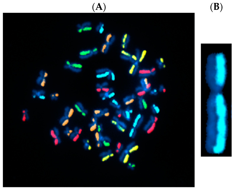
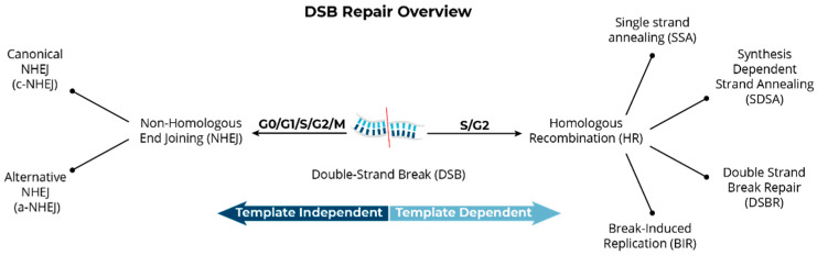
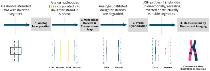
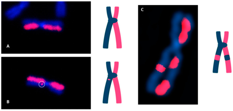
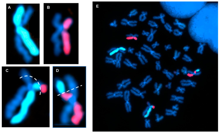
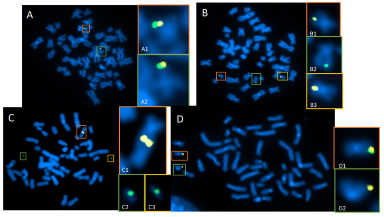
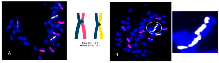
The cytogenomics-based methodology of directional genomic hybridization (dGH) enables the detection and quantification of a more comprehensive spectrum of genomic structural variants than any other approach currently available, and importantly, does so on a single-cell basis. Thus, dGH is well-suited for testing and/or validating new advancements in CRISPR-Cas9 gene editing systems. In addition to aberrations detected by traditional cytogenetic approaches, the strand specificity of dGH facilitates detection of otherwise cryptic intra-chromosomal rearrangements, specifically small inversions. As such, dGH represents a powerful, high-resolution approach for the quantitative monitoring of potentially detrimental genomic structural rearrangements resulting from exposure to agents that induce DNA double-strand breaks (DSBs), including restriction endonucleases and ionizing radiations. For intentional genome editing strategies, it is critical that any undesired effects of DSBs induced either by the editing system itself or by mis-repair with other endogenous DSBs are recognized and minimized. In this paper, we discuss the application of dGH for assessing gene editing-associated structural variants and the potential heterogeneity of such rearrangements among cells within an edited population, highlighting its relevance to personalized medicine strategies.

Keywords: DNA repair; chromosome aberrations; directional genomic hybridization; gene editing; structural variants.


Conflict of interest statement

S.M.B. and J.S.B. are cofounders and Scientific Advisory Board members of KromaTiD, Inc. E.M.C., L.K-B., H.C.S. and C.J.T. were employed by KromaTiD, Inc., at the time this work was carried out. No other interests are declared.

Figures

Figure 1
Figure 1
Directional genomic hybridization (dGH). (A) Normal, untreated human metaphase chromosome spread (peripheral blood mononuclear cells: PBMCs) illustrating strand-specific hybridization of single-stranded unique probes to the entire genome (5-color dGH SCREEN). It can be readily appreciated that, overall, there are very few structural variants in normal cells [16]. (B) Enlargement of a single chromosome 2 showing no structural rearrangements or inversions.
Figure 2
Figure 2
Overview of primary DSB repair pathways in mammalian cells. Pathway choice is determined by multiple variables, including cell type, stage of the cell cycle, genomic location of the DSB, and the availability of repair factors.
Figure 3
Figure 3
Overview of the dGH process and inversion detection. Two DSBs followed by an intra-change mis-repair (mis-rejoining) event in G1 result in an inverted segment of genomic DNA (shown within the box). 1. Analog incorporation. The genomic DNA, including the inverted segment, is replicated in the presence of the photosensitive analog nucleotides BrdU and BrdC during a single S-phase. These analogs are incorporated into the newly synthesized DNA strands and cells are arrested in the first metaphase to ensure that the analogs are incorporated solely into newly replicated daughter strands. 2. Metaphase harvest and chromosome preparation. Slides are exposed to UV light, which preferentially nicks the daughter strands at sites of analog incorporation and targets them for exonuclease degradation. After daughter-strand exonuclease degradation, chromosomes are left with original parental strands that are complementary to one another and of opposite 5′-to-3′ orientation (anti-parallel). 3. Probe hybridization. dGH single-stranded probes complementary to either one or the other single-stranded chromatid are designed against the reference sequenced genome to hybridize in a directionally specific manner. 4. Fluorescent imaging. The unique directionality of each chromatid leaves probes hybridized to a single chromatid of the chromosome, creating a fluorescently labeled “light” strand and a non-fluorescently labeled “dark” strand. Any inverted segments are readily visualized as a fluorescence pattern “switching” to the “dark” side.
Figure 4
Figure 4
dGH detection of chromosomal inversions (structural variants). (A) Image of a normal chromosome 1 with no inversion. (B) Chromosome 2 with a single, small inversion; estimated size: ~1–5 Mb. (C) Chromosome 4 with a single, large inversion; estimated size: 15–45 Mb.
Figure 5
Figure 5
dGH detection of a reciprocal translocation. (A,B) Example of a normal dGH staining pattern for chromosome 2 (A) and chromosome 11 (B), showing no translocation or inversion events. (C,D) dGH staining pattern showing a balanced translocation between homologs of chromosomes 2 and 11. Dotted lines represent estimated breakpoints. (E) Full metaphase chromosome spread of the cell containing (AD).
Figure 6
Figure 6
Targeted dGH detection of translocations in CRISPR/Cas9-edited T cells. T cells were transfected with CRISPR-Cas9 and guide RNAs for an edit site located on chromosome 19 (Chr19). Two probes (green and yellow) were designed to bracket the Chr19 edit site. (A) Cell with normal assay configuration, where yellow and green probes are co-localized on each homolog of Chr19 (A1,A2). (B) Normal homolog (B1) with normal signal pattern, and a reciprocal translocation of the green probe to an off-target chromosome (B2,B3), likely occurring at the cut site on Chr19. (C) Example of a cell containing an unbalanced translocation between both homologs of Chr19, resulting in a dicentric chromosome containing both yellow signals (C1) plus two separate acentric fragments containing the green signal (C2,C3). (D) Normal homolog (D1) with normal signal pattern, and an edit-site resection, where deletion of a large region adjacent to the edit site resulted in loss of the green signal on one Chr19 homolog (D2).
Figure 7
Figure 7
Genome editing can result in a heterogenous population of normal and abnormal cells. Human CD8+ T cells were edited with CRISPR/Cas9 ribonucleoprotein complexes. (A) Example of a structural error-free cell (with respect to the paint probes). The edited chromosome is painted yellow (2 homologs) and 3 putatively unedited chromosomes (2 homologs each) are painted pink. (B) Cell containing a complex edit-site-associated mis-repair event involving two edited homologs of chromosome 2, resulting in a dicentric chromosome (circled and enlarged), and copy number gain (Ch2). The cell is also aneuploid for chromosome 1 (trisomy).

Similar articles

Cited by

  • Development and extensive sequencing of a broadly-consented Genome in a Bottle matched tumor-normal pair.
    McDaniel JH, Patel V, Olson ND, He HJ, He Z, Cole KD, Gooden AA, Schmitt A, Sikkink K, Sedlazeck FJ, Doddapaneni H, Jhangiani SN, Muzny DM, Gingras MC, Mehta H, Behera S, Paulin LF, Hastie AR, Yu HC, Weigman V, Rojas A, Kennedy K, Remington J, Salas-González I, Sudkamp M, Wiseman K, Lajoie BR, Levy S, Jain M, Akeson S, Narzisi G, Steinsnyder Z, Reeves C, Shelton J, Kingan SB, Lambert C, Bayabyan P, Wenger AM, McLaughlin IJ, Adamson A, Kingsley C, Wescott M, Kim Y, Paten B, Park J, Violich I, Miga KH, Gardner J, McNulty B, Rosen GL, McCoy R, Brundu F, Sayyari E, Scheffler K, Truong S, Catreux S, Hannah LC, Lipson D, Benjamin H, Iremadze N, Soifer I, Krieger G, Eacker S, Wood M, Cross E, Husar G, Gross S, Vernich M, Kolmogorov M, Ahmad T, Keskus A, Bryant A, Thibaud-Nissen F, Trow J, Proszynski J, Hirschberg JW, Ryon K, Mason CE, Bhakta MS, Zachary Sanborn J, Munding EM, Wagner J, Xiao C, Liss AS, Zook JM. McDaniel JH, et al. bioRxiv [Preprint]. 2025 Jun 14:2024.09.18.613544. doi: 10.1101/2024.09.18.613544. bioRxiv. 2025. Update in: Sci Data. 2025 Jul 16;12(1):1195. doi: 10.1038/s41597-025-05438-2. PMID: 39345378 Free PMC article. Updated. Preprint.
  • Development and extensive sequencing of a broadly-consented Genome in a Bottle matched tumor-normal pair.
    McDaniel JH, Patel V, Olson ND, He HJ, He Z, Cole KD, Gooden AA, Schmitt A, Sikkink K, Sedlazeck FJ, Doddapaneni H, Jhangiani SN, Muzny DM, Gingras MC, Mehta H, Behera S, Paulin LF, Hastie AR, Yu HC, Weigman V, Rojas A, Kennedy K, Remington J, Salas-González I, Sudkamp M, Wiseman K, Lajoie BR, Levy S, Jain M, Akeson S, Narzisi G, Steinsnyder Z, Reeves C, Shelton J, Kingan SB, Lambert C, Baybayan P, Wenger AM, McLaughlin IJ, Adamson A, Kingsley C, Wescott M, Kim Y, Paten B, Park J, Violich I, Miga KH, Gardner J, McNulty B, Rosen GL, McCoy R, Brundu F, Sayyari E, Scheffler K, Truong S, Catreux S, Hannah LC, Lipson D, Benjamin H, Iremadze N, Soifer I, Krieger G, Eacker S, Wood M, Cross E, Husar G, Gross S, Vernich M, Kolmogorov M, Ahmad T, Keskus AG, Bryant A, Thibaud-Nissen F, Trow J, Proszynski J, Hirschberg JW, Ryon K, Mason CE, Bhakta MS, Sanborn JZ, Munding EM, Wagner J, Xiao C, Liss AS, Zook JM. McDaniel JH, et al. Sci Data. 2025 Jul 16;12(1):1195. doi: 10.1038/s41597-025-05438-2. Sci Data. 2025. PMID: 40670386 Free PMC article.

References

    1. Doudna J.A., Charpentier E. Genome editing. The new frontier of genome engineering with CRISPR-Cas9. Science. 2014;346:1258096. doi: 10.1126/science.1258096. - DOI - PubMed
    1. Hsu P.D., Lander E.S., Zhang F. Development and applications of CRISPR-Cas9 for genome engineering. Cell. 2014;157:1262–1278. doi: 10.1016/j.cell.2014.05.010. - DOI - PMC - PubMed
    1. Wang H., La Russa M., Qi L.S. CRISPR/Cas9 in Genome Editing and Beyond. Annu. Rev. Biochem. 2016;85:227–264. doi: 10.1146/annurev-biochem-060815-014607. - DOI - PubMed
    1. Wang J.Y., Doudna J.A. CRISPR technology: A decade of genome editing is only the beginning. Science. 2023;379:eadd8643. doi: 10.1126/science.add8643. - DOI - PubMed
    1. Hunt J.M.T., Samson C.A., Rand A.D., Sheppard H.M. Unintended CRISPR-Cas9 editing outcomes: A review of the detection and prevalence of structural variants generated by gene-editing in human cells. Hum. Genet. 2023;142:705–720. doi: 10.1007/s00439-023-02561-1. - DOI - PMC - PubMed