Review
. 2023 Dec 19;10(1):1-13.
doi: 10.3390/tomography10010001.

Advantages of Photon-Counting Detector CT in Aortic Imaging

Affiliations
Review

Advantages of Photon-Counting Detector CT in Aortic Imaging

Chiara Zanon et al. Tomography. .

Abstract

Photon-counting Computed Tomography (PCCT) is a promising imaging technique. Using detectors that count the number and energy of photons in multiple bins, PCCT offers several advantages over conventional CT, including a higher image quality, reduced contrast agent volume, radiation doses, and artifacts. Although PCCT is well established for cardiac imaging in assessing coronary artery disease, its application in aortic imaging remains limited. This review summarizes the available literature and provides an overview of the current use of PCCT for the diagnosis of aortic imaging, focusing mainly on endoleaks detection and characterization after endovascular aneurysm repair (EVAR), contrast dose volume, and radiation exposure reduction, particularly in patients with chronic kidney disease and in those requiring follow-up CT.

Keywords: CT angiography; EVAR; aortic imaging; contrast agents; dose exposure; endoleaks; photon-counting; vascular.


Conflict of interest statement

The authors declare no conflict of interest.

Figures
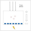
Figure 1
Figure 1
Physical principle of the Photon-counting Detector CT.
Figure 2
Figure 2
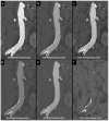
Comparison of image quality of an abdominal aortic aneurysm evaluated with photon-counting CT and standard contrast media (axial images). A thickened aortic wall related to aortitis (between solid and empty arrowhead) and intraluminal thrombotic stratification (* white asterisks) are visible at the level of the abdominal aortic aneurysm. High Resolution evaluation (Matrix 1024 × 1024) (a); Virtual Non-Contrast VCN (b); Iodine Map (c); 55 keV reconstruction (d); Pure Lumen reconstruction (e); Spectral Dual Energy Reconstruction (f). Department of Radiology, Fondazione Toscana Gabriele Monasterio, Pisa, Italy.
Figure 3
Figure 3
Comparison of image quality with photon-counting CT and standard contrast media. Abdominal aortic lumen with wall calcification axial images. High Resolution images (Matrix 1024 × 1024) with different reconstruction kernels (window level W2000, C700): 40 keV Kernel Bv48, Q4 (a); 55 keV Kernel Bv48, Q4 (b); 55 keV Kernel Bv68, Q4 (c); 70 keV Kernel Bv48, Q4 (d); Iodine map reconstruction, kernel Bv40, Q4 (e); Virtual Non-Contrast VCN reconstruction (f); 3D-Cinematic Rendering of the abdominal aorta (g). Department of Radiology, Fondazione Toscana Gabriele Monasterio, Pisa, Italy.
Figure 4
Figure 4
Comparison of image quality with photon-counting CT and standard contrast media. Abdominal aortic lumen with wall calcification coronal images. High Resolution images (Matrix 1024 × 1024) with different reconstruction kernels (window level W2000, C700): 40 keV Kernel Bv48, Q4 (a); 55 keV Kernel Bv48, Q4 (b); 55 keV Kernel Bv68, Q4 (c); 70 keV Kernel Bv48, Q4 (d); Iodine map reconstruction, kernel Bv40, Q4 (e); Virtual Non Contrast VCN reconstruction (f). Department of Radiology, Fondazione Toscana Gabriele Monasterio, Pisa, Italy.
Figure 5
Figure 5
Aortic angiographic evaluation of the abdominal aortic aneurysm evaluated with photon-counting CT and standard contrast media (the same example of Figure 2, coronal view). The aortic wall is indicated between solid and empty arrowhead. 3D-Volume Rendering representation (a); High-Resolution coronal evaluation, 55 keV, Kernel Bv68 Q4 Matrix 1024 (b); Spectral Dual Energy coronal Reconstruction (c). Department of Radiology, Fondazione Toscana Gabriele Monasterio, Pisa, Italy.

Similar articles

Cited by

References

    1. Meloni A., Frijia F., Panetta D., Degiorgi G., De Gori C., Maffei E., Clemente A., Positano V., Cademartiri F. Photon-Counting Computed Tomography (PCCT): Technical Background and Cardio-Vascular Applications. Diagnostics. 2023;13:645. doi: 10.3390/diagnostics13040645. - DOI - PMC - PubMed
    1. Rubin G.D., Leipsic J., Schoepf U.J., Fleischmann D., Napel S. CT Angiography after 20 Years: A Transformation in Cardiovascular Disease Characterization Continues to Advance. Radiology. 2014;271:633–652. doi: 10.1148/radiol.14132232. - DOI - PMC - PubMed
    1. Pepe A., Crimì F., Vernuccio F., Cabrelle G., Lupi A., Zanon C., Gambato S., Perazzolo A., Quaia E. Medical Radiology: Current Progress. Diagnostics. 2023;13:2439. doi: 10.3390/diagnostics13142439. - DOI - PMC - PubMed
    1. Manghat N., Morgan-Hughes G., Roobottom C. Multi-detector row computed tomography: Imaging in acute aortic syndrome. Clin. Radiol. 2005;60:1256–1267. doi: 10.1016/j.crad.2005.06.011. - DOI - PubMed
    1. Schlett C.L., Pursnani A., Marcus R.P., Truong Q.A., Hoffmann U. The Use of Coronary CT Angiography for the Evaluation of Chest Pain. Cardiol. Rev. 2014;22:117–127. doi: 10.1097/CRD.0000000000000015. - DOI - PubMed

MeSH terms

LinkOut - more resources