Review
. 2023 Dec 19;16(1):2.
doi: 10.3390/toxins16010002.

A Review of Rattlesnake Venoms

Affiliations
Review

A Review of Rattlesnake Venoms

Phuc Phan et al. Toxins (Basel). .

Abstract

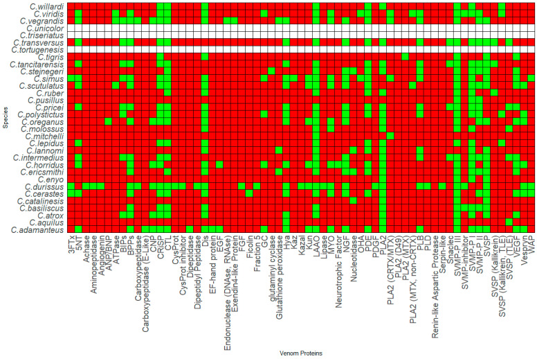
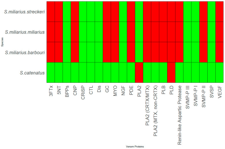
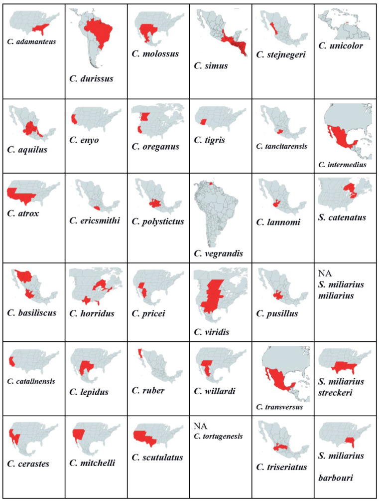
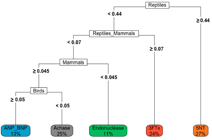
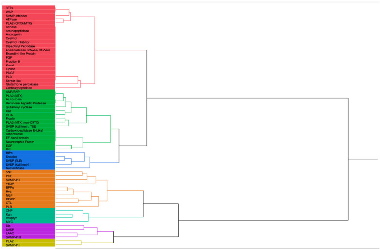
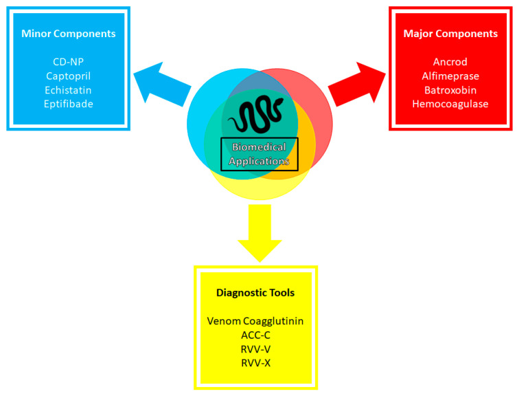
Venom components are invaluable in biomedical research owing to their specificity and potency. Many of these components exist in two genera of rattlesnakes, Crotalus and Sistrurus, with high toxicity and proteolytic activity variation. This review focuses on venom components within rattlesnakes, and offers a comparison and itemized list of factors dictating venom composition, as well as presenting their known characteristics, activities, and significant applications in biosciences. There are 64 families and subfamilies of proteins present in Crotalus and Sistrurus venom. Snake venom serine proteases (SVSP), snake venom metalloproteases (SVMP), and phospholipases A2 (PLA2) are the standard components in Crotalus and Sistrurus venom. Through this review, we highlight gaps in the knowledge of rattlesnake venom; there needs to be more information on the venom composition of three Crotalus species and one Sistrurus subspecies. We discuss the activity and importance of both major and minor components in biomedical research and drug development.

Keywords: COVID-19; Crotalus; Sistrurus; biomedical application; toxin; venom composition.


Conflict of interest statement

The authors declare no conflict of interest.

Figures

Figure 1
Figure 1
Presence-absence analysis of the genus Crotalus venom composition. The color of the box represents the presence (green), absence (red), and no information (white). The graph was generated using R. Abbreviations: Three-finger toxin (3FTx), 5′-nucleotidase (5′-NT), Acetylcholinesterase (Achase), Natriuretic peptide type A (ANP), Adenosine triphosphatase (ATPase), Bradykinin inhibitory peptide (BIP), Natriuretic peptide type B (BNP), Bradykinin potentiate peptide (BPP), C-type Lectins (CTL), Natriuretic peptide type C (CNP), Cysteine Protease (CYSPROT), Cysteine-rich secretory protein (CRiSP), Crotoxin (CRTX), Disintegrin (DIS), Epidermal growth factor (EGF), Fibroblast growth factor (FGF), Guanylyl cyclase (GC), Hyaluronidase (HYA), Kazal-type inhibitor (KAZAL), Kunitz-type inhibitor (KUN), L-amino acid oxidase (LAAO), Mojave toxin (MTX), Myotoxin (MYO), Nerve growth factor (NGF), Ohanin (OHA), Phosphodiesterase (PDE), Platelet-derived growth factor (PDGF), Phospholipase A2 (PLA2), Phospholipase B (PLB), Phospholipase D (PLD), Snake venom metalloprotease (SVMP), Snake venom serine protease (SVSP), Snake C-type Lectin (SNACLEC), Thrombin-like enzyme (TLE), Vascular endothelial growth factor (VEGF), Waparin (WAP).
Figure 2
Figure 2
Presence-absence analysis of the genus Sistrurus venom composition. The color of the box represents the presence (green), absence (red), and no information (white). The graph was generated using R. Abbreviations: Three-finger toxin (3FTx), 5′-nucleotidase (5′-NT), Bradykinin potentiating peptide (BPP), C-type Lectins (CTL), Natriuretic peptide type C (CNP), Cysteine-rich secretory protein (CRiSP), Crotoxin (CRTX), Disintegrin (DIS), Guanylyl Cyclase (GC), Hyaluronidase (HYA), L-amino acid oxidase (LAAO), Myotoxin (MYO), Nerve growth factor (NGF), Phosphodiesterase (PDE), Phospholipase A2 (PLA2), Phospholipase B (PLB), Snake venom metalloproteinase (SVMP), Snake venom serine protease (SVSP), Vascular endothelial growth factor (VEGF).
Figure 3
Figure 3
Range maps for each of the 36 Crotalus species. The red color in the map denotes the range of the species.
Figure 4
Figure 4
Decision tree demonstrating the role diet can play in the venom composition within rattlesnakes. We want to emphasize that the patterns demonstrated in this study in the venom composition are limited as most of the studies did not specify the rattlesnake’s source, age, and diet composition at the time of venom collection.
Figure 5
Figure 5
This dendrogram was created via the hierarchical clustering of proteins in rattlesnake venom by habitat type and distribution patterns. The proteins in red are most likely to be found in Central America. The proteins highlighted in green are most likely found in SW/S/W US and northern and southern Mexico. The proteins in blue are most likely to be found in SW/S/W US and south and western Mexico. The proteins in orange color are found throughout the range of rattlesnakes but are highly common in SW/S/W US and southern Mexico. Proteins highlighted by ocean green are mainly found in SE/SW/S/W US and northern Mexico. The proteins in purple color are present in high quantity throughout the rattlesnake range. The proteins in light green color are found in high abundance in central and southern Mexico. We want to emphasize that the patterns demonstrated in this study in the venom composition are limited as most of the studies did not specify the rattlesnake’s source, age, and diet composition at the time of venom collection.
Figure 6
Figure 6
Biomedical applications of snake venoms. The graph demonstrates notable applications of snake venoms in biomedical research. Applications are separated into three groups: therapeutic drugs based on major components and minor components of snake venom, and diagnostic tools that are used in biomedical diagnosis and analysis.

References

    1. Fry B.G., Wüster W., Kini R.M., Brusic V., Khan A., Venkataraman D., Rooney A.P. Molecular evolution and phylogeny of elapid snake venom three-finger toxins. J. Mol. Evol. 2003;57:110–129. doi: 10.1007/s00239-003-2461-2. - DOI - PubMed
    1. Tasoulis T., Isbister G.K. A Review and Database of Snake Venom Proteomes. Toxins. 2017;9:290. doi: 10.3390/toxins9090290. - DOI - PMC - PubMed
    1. Assakura M.T., Salomao M.G., Puorto G., Mandelbaum F.R. Hemorrhagic, fibrinogenolytic and edema-forming activities of the venom of the colubrid snake Philodryas olfersii (green snake) Toxicon. 1992;30:427–438. doi: 10.1016/0041-0101(92)90539-H. - DOI - PubMed
    1. Daltry J.C., Wuster W., Thorpe R.S. Diet and snake venom evolution. Nature. 1996;379:537–540. doi: 10.1038/379537a0. - DOI - PubMed
    1. Fry B.G., Wickramaratna J.C., Hodgson W.C., Alewood P.F., Kini R., Ho H., Wüster W. Electrospray liquid chromatography/mass spectrometry fingerprinting of Acanthophis (death adder) venoms: Taxonomic and toxinological implications. Rapid Commun. Mass. Spectrom. 2002;16:600–608. doi: 10.1002/rcm.613. - DOI - PubMed

LinkOut - more resources