. 2024 Jan 20;29(2):521.
doi: 10.3390/molecules29020521.

Molecular Characterization of the Recombinant Ig1 Axl Receptor Domain: An Intriguing Bait for Screening in Drug Discovery

Affiliations

Molecular Characterization of the Recombinant Ig1 Axl Receptor Domain: An Intriguing Bait for Screening in Drug Discovery

Rossella Di Stasi et al. Molecules. .

Abstract

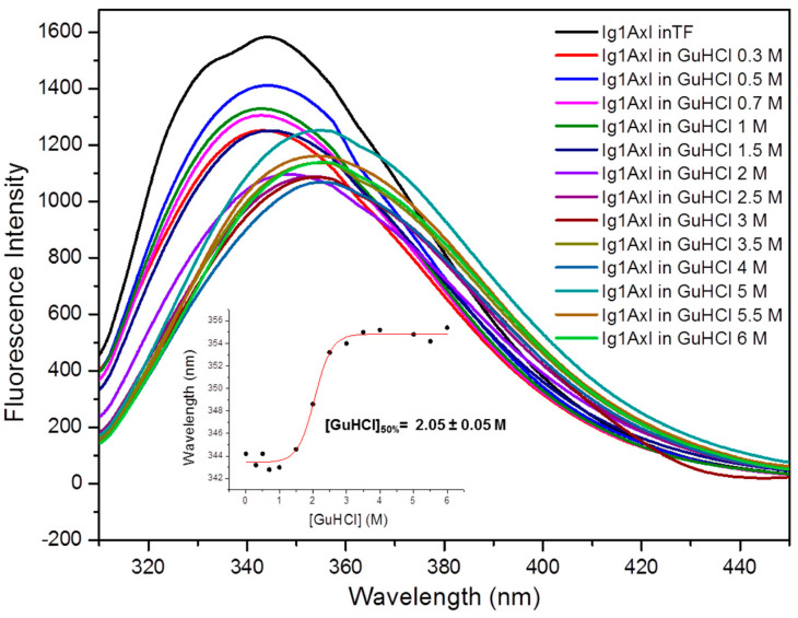
Axl receptor tyrosine kinase and its ligand Gas6 regulate several biological processes and are involved in both the onset and progression of tumor malignancies and autoimmune diseases. Based on its key role in these settings, Axl is considered a promising target for the development of molecules with therapeutic and diagnostic purposes. In this paper, we describe the molecular characterization of the recombinant Ig1 domain of Axl (Ig1 Axl) and its biochemical properties. For the first time, an exhaustive spectroscopic characterization of the recombinant protein through circular dichroism and fluorescence studies is also reported, as well as a binding analysis to its natural ligand Gas6, paving the way for the use of recombinant Ig1 Axl as a bait in drug discovery screening procedures aimed at the identification of novel and specific binders targeting the Axl receptor.

Keywords: Axl receptor; circular dichroism; drug discovery; fluorescence spectroscopy; iodoacetamide; protein refolding; recombinant Ig1 domain; trypsin.


Conflict of interest statement

The authors declare no conflicts of interest.

Figures

Figure 1
Figure 1
SDS-PAGE analysis of the (a) recombinant expression of the Ig1 Axl protein in the BL21-Gold(DE3)pLysS E. coli strain; (b) Ig1 Axl TEV cleavage; (c) recombinant Ig1 Axl concentrated in Amicon Ultra centrifugal filters ultracel after the size-exclusion chromatography purification step. Lane 1 of the gel (a) E. coli not induced crude extract in Tris-HCl 50 mM/NaCl 150 mM, pH = 7; Lanes 2 and 3: E. coli-induced crude extract supernatant in Tris-HCl 50 mM/NaCl 150 mM, pH = 7 (5 and 10 μL, respectively); Lane 4: E. coli-induced crude extract pellet in Tris-HCl 50 mM/NaCl 150 mM, pH = 7 (10 μL). In (ac), Ig1 Axl is circled in the magenta ovals. M, pre-stained protein molecular weight marker (11–245 KDa). (d) the LC profile from the LC-MS ESI-ToF analysis of pure recombinant Ig1 Axl. The deconvoluted spectrum and the value of the average mass are reported in (e).
Figure 2
Figure 2
Analysis with ESI-ToF mass spectrometry of the recombinant Ig1 Axl domain after (A) incubation at 60 °C for 90 min and alkylation with iodoacetamide at RT for 30 min and (B) incubation with tris-carboxyethylphosphine (TCEP) at 60 °C for 90 min and alkylation with iodoacetamide at RT for 30 min. In the inset of panels, (A,B) report the deconvoluted mass spectra showing the experimental average mass values of the species. ¥ Ig1 Axl, MWth (av): 12,036.288 Da; * dialkylated Ig1 Axl, MWth (av): 12,152.388 Da; $ monoalkylated MWth (av): 12,095.338 Da; # reduced Ig1 Axl, MWth (av): 12,038.288 Da.
Figure 3
Figure 3
Analysis using LC-ESI-ToF mass spectrometry of the Ig1 Axl trypsin digestion mixture after 3 h of incubation with trypsin at 37 °C. (A) Chromatographic profile revealed by registering the total ionic current. (B) ESI-ToF spectra of the peptide fragments obtained.
Figure 4
Figure 4
Ig1 Axl trypsin proteolytic digestion mixture analyzed with LC-MS after 3 h of incubation at 37 °C. (A) Ig1 Axl protein sequence in which the trypsin cleavage sites are underlined and the corresponding residue is numbered. The numbering refers to the human Axl receptor protein sequence annotated in the UniProt Database (entry P30530). The cysteine residues are in bold red. (B) List of trypsin-derived peptides of recombinant Ig1 Axl. a Amino acid sequences of the peptide fragments obtained from recombinant Ig1 Axl after trypsin digestion; b experimental (exp) and theoretical (th) molecular weight (MW) of the LC-MS-identified peptide fragments; c chromatographic retention time (TR) of the identified peptide fragments. The peptide fragments 56C–R71 and 104I–E136 reported in panel (B) correspond to a peptide fragment harboring the cysteine residues (56C and 117C) in the oxidized state joined with the disulfide bridge.
Figure 5
Figure 5
Analysis with LC-ESI-ToF mass spectrometry of the Ig1 Axl biotinylation reaction mixture after 1 h of incubation with NHS-dPEG®4-biotin at RT. (A) RP-HPLC chromatographic profile revealed by registering the absorbance at 210 nm. (B) ESI-ToF mass spectrum of the protein species eluted at 19.280 min. (C) Deconvoluted mass spectrum. * residual unlabeled Ig1 Axl (MWth: 12,036.288 Da); # Ig1 Axl labeled with dPEG®4-biotin (MWth: 12,509.868 Da); $ trace amount of Ig1 Axl labeled with two dPEG®4-biotin units (MWth: 12,983.448 Da). The minor peaks eluted at 13.137 min and 14.457 min correspond to the reactive probe NHS-dPEG®4-biotin (§) and to the hydrolysis product of the probe dPEG®4-biotin (¤).
Figure 6
Figure 6
Binding analysis of recombinant Ig1 Axl to Gas6 LG1-LG2 with bio-layer interferometry. (A) Binding traces of biotinylated Ig1 Axl (10 nM) incubated with the indicated concentrations of Gas6 LG1-LG2. (B) Steady-state analysis derived by the fitting of the association and dissociation curves performed using the global algorithm and the 1:1 binding model.
Figure 7
Figure 7
Recombinant Ig1 Axl thermal denaturation analysis using CD spectroscopy. The figure shows the superimposition of the CD spectra acquired in the temperature range of 10–90 °C. The data were collected over five averaged scans. The inset reports the thermal denaturation curve as the result of a non-linear regression analysis (R2 = 0.99) obtained by plotting the temperature dependence of the ellipticity values registered at 202 nm ([θ]202).
Figure 8
Figure 8
Ig1 Axl analysis created with steady-state fluorescence spectroscopy. The figure reports the superimposition of the Ig1 Axl emission spectrum recorded at 20 °C in 10 mM phosphate buffer (TF) at pH 7 (black curve) and the protein spectra acquired in the presence of increasing concentrations of GuHCl (colored curves from 0.3 to 6 M GuHCl). The inset shows the sigmoidal curve obtained by plotting the wavelength shift of the maximum fluorescence intensity as a function of GuHCl concentration, which represents the result of the non-linear regression analysis (R2 = 0.99).

References

    1. Tsou W.I., Nguyen K.Q., Calarese D.A., Garforth S.J., Antes A.L., Smirnov S.V., Almo S.C., Birge R.B., Kotenko S.V. Receptor tyrosine kinases, TYRO3, AXL, and MER, demonstrate distinct patterns and complex regulation of ligand-induced activation. J. Biol. Chem. 2014;289:25750–25763. doi: 10.1074/jbc.M114.569020. - DOI - PMC - PubMed
    1. Lew E.D., Oh J., Burrola P.G., Lax I., Zagorska A., Traves P.G., Schlessinger J., Lemke G. Differential TAM receptor-ligand-phospholipid interactions delimit differential TAM bioactivities. eLife. 2014;3:e03385. doi: 10.7554/eLife.03385. - DOI - PMC - PubMed
    1. Wium M., Paccez J.D., Zerbini L.F. The Dual Role of TAM Receptors in Autoimmune Diseases and Cancer: An Overview. Cells. 2018;7:166. doi: 10.3390/cells7100166. - DOI - PMC - PubMed
    1. Bellan M., Pirisi M., Sainaghi P.P. The Gas6/TAM System and Multiple Sclerosis. Int. J. Mol. Sci. 2016;17:1807. doi: 10.3390/ijms17111807. - DOI - PMC - PubMed
    1. Meertens L., Labeau A., Dejarnac O., Cipriani S., Sinigaglia L., Bonnet-Madin L., Le Charpentier T., Hafirassou M.L., Zamborlini A., Cao-Lormeau V.M., et al. Axl Mediates ZIKA Virus Entry in Human Glial Cells and Modulates Innate Immune Responses. Cell Rep. 2017;18:324–333. doi: 10.1016/j.celrep.2016.12.045. - DOI - PubMed