. 2024 Jan 22;16(2):301.
doi: 10.3390/polym16020301.

Water-Soluble Star Polymer as a Potential Photoactivated Nanotool for Lysozyme Degradation

Affiliations

Water-Soluble Star Polymer as a Potential Photoactivated Nanotool for Lysozyme Degradation

Lidia Mezzina et al. Polymers (Basel). .

Abstract

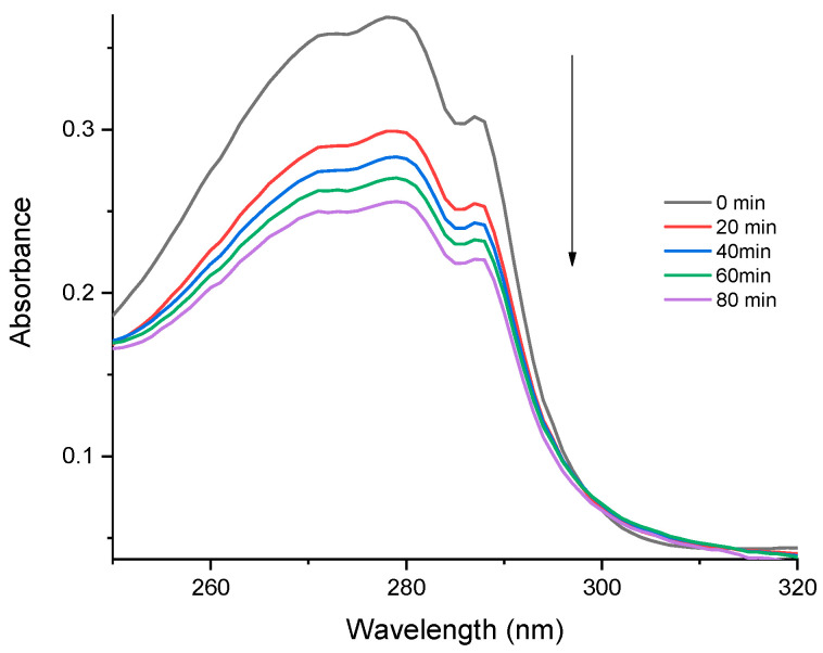
The development of nanotools for chemical sensing and macromolecular modifications is a new challenge in the biomedical field, with emphasis on artificial peptidases designed to cleave peptide bonds at specific sites. In this landscape, metal porphyrins are attractive due to their ability to form stable complexes with amino acids and to generate reactive oxygen species when irradiated by light of appropriate wavelengths. The issues of hydrophobic behavior and aggregation in aqueous environments of porphyrins can be solved by using its PEGylated derivatives. This work proposes the design of an artificial photo-protease agent based on a PEGylated mercury porphyrin, able to form a stable complex with l-Tryptophan, an amino acid present also in the lysozyme structure (a well-known protein model). The sensing and photodegradation features of PEGylated mercury porphyrin were exploited to detect and degrade both l-Trp and lysozyme using ROS, generated under green (532 nm) and red (650 nm) light lasers. The obtained system (Star3600_Hg) and its behavior as a photo-protease agent were studied by means of several spectroscopies (UV-Vis, fluorescence and circular dichroism), and MALDI-TOF mass spectrometry, showing the cleavage of lysozyme and the appearance of several short-chain residues. The approach of this study paves the way for potential applications in theranostics and targeted bio-medical therapies.

Keywords: l-tryptophan; lysozyme; photo-degradation; porphyrin; protein degradation.


Conflict of interest statement

The authors declare no conflicts of interest.

Figures

Figure 1
Figure 1
MALDI-TOF mass spectra of Star3600_2H (a) and Star3600_Hg (b).
Scheme 1
Scheme 1
Exemplificative scheme of the synthesis of Star3600_Me.
Figure 2
Figure 2
UV-Vis (continuous lines) and fluorescence (dashed lines) spectra of water solutions of Star3600_2H (red lines, λexc = 430 nm), Star3600_Zn (green lines, λexc = 435 nm) and Star3600_Hg (blue lines, λexc = 440 nm. Spectrum magnified ×25).
Figure 3
Figure 3
(a) Singlet oxygen production mechanism scheme and (b) RNO degradation mechanism mediated by the 1O2–imidazole system.
Figure 4
Figure 4
Upper: circular dichroism spectra of aqueous solutions of pristine Star3600_Hg (black line) and of Star3600_Er (green), Star3600_Rh (cyan), Star3600_Sn (magenta), Star3600_Zn (blue) and Star3600_Hg (red line) when mixed with l-Trp (molar ratio 1:100). Bottom: relative UV-Vis spectra in the Soret band region.
Figure 5
Figure 5
UV-vis (continuous lines) and fluorescence (dashed lines) spectra of aqueous solutions of Star3600_Hg (black line) and a mixture of Star3600_Hg and l-Trp in molar ratio 1:100 freshly prepared (red line) and after 10 and 120 min (blue and green lines, respectively).
Figure 6
Figure 6
RNO degradation mechanism mediated by the 1O2l-Trp system.
Figure 7
Figure 7
UV-vis spectra of the mixture of 5 µM aqueous solution of Star3600_Hg and l-TRP (ratio 1:100) when exposed to a red laser (650 nm) irradiation.
Figure 8
Figure 8
UV-Vis (continuous lines) and fluorescence (dashed lines) spectra (λexc = 440 nm) of Star3600_Hg/LSZ complex. The black line is an aqueous solution (5 µM) of Star3600_Hg, and red and green lines are the aqueous solution mixture of Star3600_Hg with LSZ (molar ratio 1:1) freshly prepared and after 2 days, respectively.
Figure 9
Figure 9
Fluorescence spectra (λexc = 290 nm) of the aqueous solution of Star3600_Hg and LSZ mixture (both 5 µM) under (a) green (532 nm) and (b) red (650 nm) laser irradiation.
Figure 10
Figure 10
(a) CD spectra of aqueous solutions of LSZ (blue line), Star3600_Hg and LSZ (both 5 µM) mixture freshly prepared (black line), and after 40 (green dash dot line) and 80 (green solid line) minutes under green laser irradiation; (b) magnification of the range 400–490 nm and (c) the related UV-vis spectra.
Figure 11
Figure 11
MALDI-TOF mass spectra of pristine lysozyme (black line) and Star3600_Hg-LSZ solution after 80 min of laser irradiation (magenta line). The symbols are explained in Table 3.
Figure 12
Figure 12
Aminoacidic sequence of lysozyme. W indicates l-Trp residues.
Figure 13
Figure 13
Schematic representation of the Star 3600_Hg photo-induced proteolysis mechanism.

References

    1. Kumar R., Singh R., Hui D., Feo L., Fraternali F. Graphene as biomedical sensing element: State of art review and potential engineering applications. Compos. Part B Eng. 2018;134:193–206. doi: 10.1016/j.compositesb.2017.09.049. - DOI
    1. Hegg E.L., Burstyn J.N. Toward the development of metal-based synthetic nucleases and peptidases: A rationale and progress report in applying the principles of coordination chemistry. Coord. Chem. Rev. 1998;173:133–165. doi: 10.1016/S0010-8545(98)00157-X. - DOI
    1. Buranaprapuk A., Kumar C.V., Jockusch S., Turro N.J. Photochemical Protein Scissors: Role of Aromatic Residues on the Binding Affinity and Photocleavage Efficiency of Pyrenyl Peptides. Tetrahedron. 2000;56:7019–7025. doi: 10.1016/S0040-4020(00)00525-1. - DOI
    1. Chowdhury N., Dutta S., Dasgupta S., Singh N.D.P., Baidya M., Ghosh S.K. Synthesis, photophysical, photochemical, DNA cleavage/binding and cytotoxic properties of pyrene oxime ester conjugates. Photochem. Photobiol. Sci. 2012;11:1239–1250. doi: 10.1039/c2pp25033k. - DOI - PubMed
    1. Vera C.C., Borsarelli C.D. Photo-induced protein modifications: A range of biological consequences and applications. Biophys. Rev. 2023;15:569–576. doi: 10.1007/s12551-023-01081-6. - DOI - PMC - PubMed

LinkOut - more resources