Case Reports
. 2024 Jan 26;44(2):56.
doi: 10.1007/s10875-024-01653-5.

Impaired STING Activation Due to a Variant in the E3 Ubiquitin Ligase AMFR in a Patient with Severe VZV Infection and Hemophagocytic Lymphohistiocytosis

Affiliations
Case Reports

Impaired STING Activation Due to a Variant in the E3 Ubiquitin Ligase AMFR in a Patient with Severe VZV Infection and Hemophagocytic Lymphohistiocytosis

Michelle Mølgaard Thomsen et al. J Clin Immunol. .

Abstract

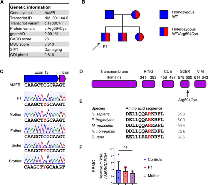
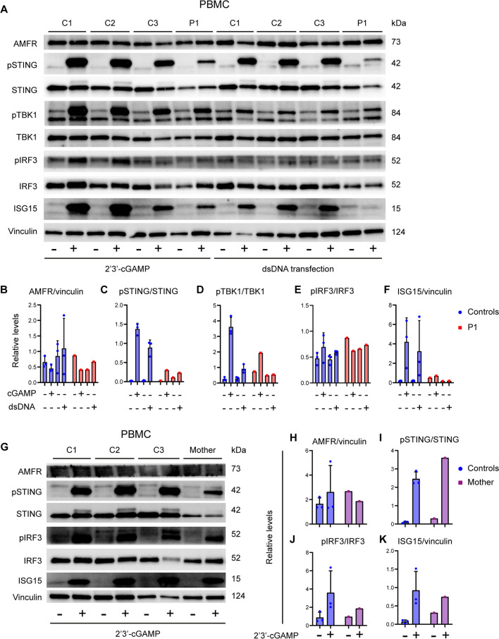
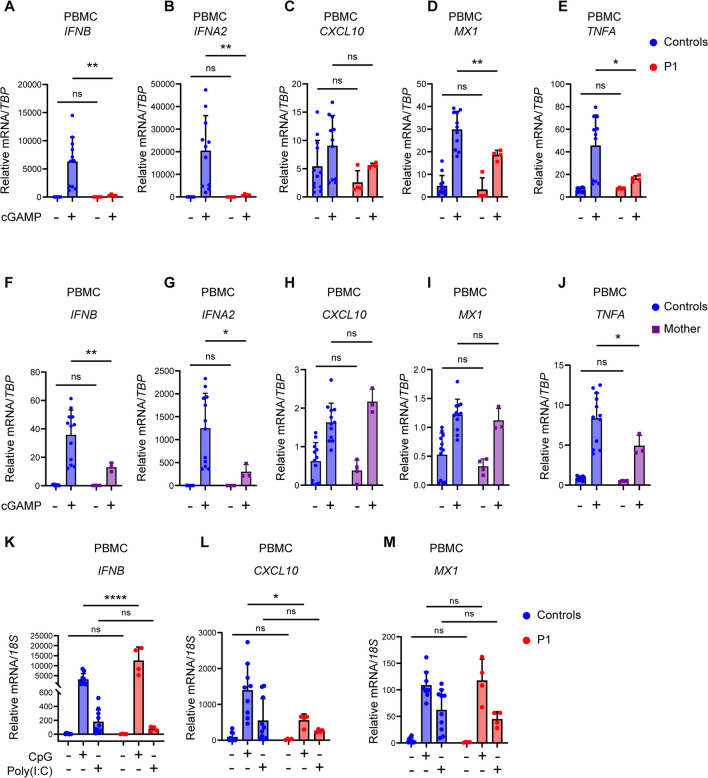
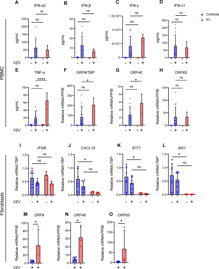
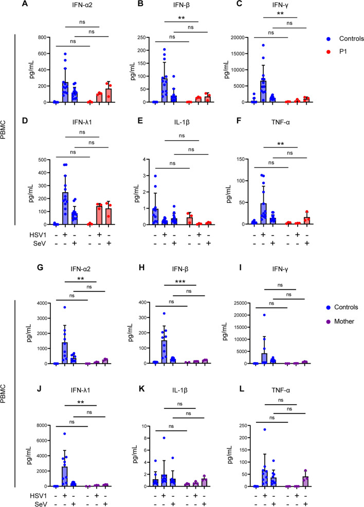
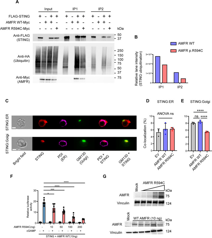
Varicella zoster virus (VZV) is a neurotropic alphaherpesvirus exclusively infecting humans, causing two distinct pathologies: varicella (chickenpox) upon primary infection and herpes zoster (shingles) following reactivation. In susceptible individuals, VZV can give rise to more severe clinical manifestations, including disseminated infection, pneumonitis, encephalitis, and vasculopathy with stroke. Here, we describe a 3-year-old boy in whom varicella followed a complicated course with thrombocytopenia, hemorrhagic and necrotic lesions, pneumonitis, and intermittent encephalopathy. Hemophagocytic lymphohistiocytosis (HLH) was strongly suspected and as the condition deteriorated, HLH therapy was initiated. Although the clinical condition improved, longstanding hemophagocytosis followed despite therapy. We found that the patient carries a rare monoallelic variant in autocrine motility factor receptor (AMFR), encoding a ubiquitin ligase involved in innate cytosolic DNA sensing and interferon (IFN) production through the cyclic GMP-AMP synthase-stimulator of IFN genes (cGAS-STING) pathway. Peripheral blood mononuclear cells (PBMCs) from the patient exhibited impaired signaling downstream of STING in response dsDNA and 2'3'-cGAMP, agonists of cGAS and STING, respectively, and fibroblasts from the patient showed impaired type I IFN responses and significantly increased VZV replication. Overexpression of the variant AMFR R594C resulted in decreased K27-linked STING ubiquitination compared to WT AMFR. Moreover, ImageStream technology revealed reduced STING trafficking from ER to Golgi in cells expressing the patient AMFR R594C variant. This was supported by a dose-dependent dominant negative effect of expression of the patient AMFR variant as measured by IFN-β reporter gene assay. Finally, lentiviral transduction with WT AMFR partially reconstituted 2'3'-cGAMP-induced STING-mediated signaling and ISG expression in patient PBMCs. This work links defective AMFR-STING signaling to severe VZV disease and hyperinflammation and suggests a direct role for cGAS-STING in the control of viral infections in humans. In conclusion, we describe a novel genetic etiology of severe VZV disease in childhood, also representing the first inborn error of immunity related to a defect in the cGAS-STING pathway.

Keywords: AMFR; HLH; ISG; Interferon; STING; Ubiquitin ligase; VZV.


Conflict of interest statement

The authors declare no competing interests.

Figures

Fig. 1
Fig. 1
Identification of a rare genetic variant in AMFR in a patient with severe VZV infection and HLH. A Characteristics of the monoallelic AMFR variant. B Pedigree showing heterozygous inheritance of the AMFR R594C variant inherited from the mother. C Sanger sequencing confirming the presence of the AMFR R594C variant in the patient (P1), the patient’s mother, and siblings. D Protein structure of AMFR with localization of the R594C variant in the G2BR domain. E Protein alignment showing conservation of Arginine at position 594 in AMFR across different species. F AMFR mRNA levels were measured using RT-qPCR of whole-cell lysates of PBMCs from healthy controls, P1, and his mother. Statistics was done by ordinary 1-way ANOVA. not significant (ns). CADD, combined annotation dependent depletion; MSC, mutation significance cutoff; SIFT, sorting intolerant from tolerant; gnomAD, the genome aggregation database; GDI, Gene Damage Index; RING, really interesting new gene; CUE, coupling of ubiquitin conjugation to ER degradation; G2BR, Ube2g2-binding region; VIM, p97/VCP-interacting motif
Fig. 2
Fig. 2
Reduced STING signaling and ISG responses in patient cells. A PBMCs from the patient and three healthy controls (C1–C3) were stimulated with 100 µg/mL of 2′3′-cGAMP (A) or 2 µg/mL transfected dsDNA for 3 h and lysates were subjected to western blotting for the expression levels of pSTING, STING, pTBK1, TBK1, pIRF3, IRF3, ISG15, and vinculin (loading control) B–F Quantification of the intensity of the western blot bands in (B). G PBMCs from the mother and three healthy controls were stimulated with 100 µg/mL of 2′3′-cGAMP for 3 h. Lysates were subjected to western blotting for the protein expression of pSTING, STING, pIRF3, IRF3, ISG15, AMFR, and vinculin (loading control). H–K Quantification of intensity of the western blot bands in (G)
Fig. 3
Fig. 3
Impaired cGAMP-induced responses and preserved TLR3 and TLR9 signaling in patient cells. PBMC from healthy controls and P1 (A–E), or his mother (F–J), were stimulated with 100 µg/mL 2′3′-cGAMP for 6 h. Total RNA was isolated and mRNA levels of IFNB, IFNA2, CXCL10, MX1, and TNFA were measured relative to TBP. K–M PBMCs from P1 and three healthy controls were stimulated with poly(I:C) (50 µg/mL) or CpG ODN2395 (5 µg/mL) for 6h, after which total RNA was isolated and levels of IFNB1, CXCL10, and MX1 were measured relative to 18S mRNA. Statistics: 2-way ANOVA with Sidak’s multiple comparisons test. Ns, non-significant; *p ≤ 0.05, **p < 0.01, ****p ≤ 0.0001
Fig. 4
Fig. 4
Antiviral and proinflammatory immune responses and viral replication in patient cells infected with cell-free VZV. A–H PBMCs from P1 and healthy controls were infected with cell-free VZV rOka vaccine-strain (VZV rOka), MOI 0.2 for 24 h. A–F Supernatants were examined for the level of IFN-α2, IFN-β, IFN-λ1, IFN-γ, and TNF-α using Mesoscale U-plex assays. F–H VZV infected PBMCs were lysed, and RNA was purified and subjected to RT-qPCR for the expression of VZV open reading frame gene (ORF)9, ORF40, and ORF63. ORF mRNA was normalized to TBP levels, and statistics were calculated using a 2-way ANOVA and Sidak’s multiple comparisons test. The experiment is representative of three independent experiments. I–O Primary fibroblasts of P1 and two healthy controls were infected with VZV rOka at MOI 1. At 48 hpi, the cells were lysed for RT-qPCR of viral ORF expression and ISG mRNA. I–L mRNA levels of IFNB, CXCL10, IFIT1, and MX1 were normalized to TBP. M–O ORF mRNA levels were normalized to PPIB. Statistics: Mann–Whitney U-test. Statistics: 2-way ANOVA and Sidak’s multiple comparisons test. Abbreviations: Ns, non-significant; *p < 0.05, ****p ≤ 0.0001
Fig. 5
Fig. 5
Induction of IFNs and proinflammatory cytokines in patient PBMCs in response to HSV1 and SeV infection. A–L PBMCs from P1 A–F or the mother G–L were infected with HSV1 (MOI 3) or SeV (50 HAU/500,000 cells), and supernatants harvested 24 h post-infection, and levels of IFN-α2, IFN-β, IFN-λ1, IFN-γ, IL-1β, and TNF-α were measured using Mesoscale U-plex assays. Statistical significance was calculated using a 2-way ANOVA and Sidak’s multiple comparisons test. Ns, non-significant; ** ≤ 0.01. The experiments were performed twice
Fig. 6
Fig. 6
Decreased STING ubiquitination in cells expressing the patient AMFR R594C variant. A HEK293T cells were transfected with 1 µg of FLAG-STING, HA-K27 ubiquitin, and AMFR-Myc-WT or AMFR-Myc-R594C for 24 h, after which STING was immunoprecipitated twice with anti-FLAG-M2 beads. Lysates were immunoblotted for expression of FLAG-STING, HA-(K27) ubiquitin, and Myc-AMFR. B HA-ubiquitin expression was quantified in ImageLab (Bio-Rad, USA) by measuring the total lane intensity of the HA-ubiquitin smear for both AMFR-WT and AMFR-R594C mutant expressing cells in IP1 and IP2. The experiments are representative of three independent experiments. C–D HEK293T cells were transfected as indicated and probed with mouse anti-STING and anti-PDI (ER marker) or anti-GM130 (Golgi marker). Cells were analyzed by ImageStream. Panel C shows representative images, and panels D and E show quantification of STING-ER colocalization and STING-Golgi colocalization from three independent experiments of STING-ER co-localization F, G HEK 293T cells with stable STING expression were transfected with 10, 50, 100, 200 ng AMFR R594C or empty vector, IFNB1 promoter luciferase reporter, and β-actin Renilla reporter. F Eighteen hours after transfection, the cells were stimulated with 2′3′-cGAMP (50 mg/mL) for 8 h. G Cells were lysed for western blotting for AMFR and vinculin (loading control). Statistics were calculated with a 2-way ANOVA and Dunnett’s multiple comparisons test. *p<0.05; ***p < 0.001; ****p < 0.0001. The experiments were performed twice
Fig. 7
Fig. 7
Reconstitution of patient PBMCs with WT AMFR restores cGAS-STING signaling and ISG responses. A Patient and control PBMCs were stimulated with 1.5 µg/mL PHA for 72 h and then transduced by VSV-G lentiviral vectors to express AMFR WT or GFP (control) (MOI 10). After 72 h, the cells were stimulated with 2′3′-cGAMP for 3 h, and cell lysates were analyzed by immunoblotting for AMFR, pSTING, STING, pTBK1, TBK1, pIRF3, IRF3, ISG15, GFP, and vinculin (loading control). (B–F) Quantification of the Western blot bands in (A)

References

    1. Zerboni L, Sen N, Oliver SL, Arvin AM. Molecular mechanisms of varicella zoster virus pathogenesis. Nat Rev Microbiol. 2014;12(3):197–210. doi: 10.1038/nrmicro3215. - DOI - PMC - PubMed
    1. Kennedy PGE, Gershon AA. Clinical features of Varicella-Zoster virus infection. Viruses. 2018;10(11):609. doi: 10.3390/v10110609. - DOI - PMC - PubMed
    1. Nagel MA, Niemeyer CS, Bubak AN. Central nervous system infections produced by varicella zoster virus. Curr Opin Infect Dis. 2020;33(3):273–278. doi: 10.1097/QCO.0000000000000647. - DOI - PubMed
    1. Bozzola E, Tozzi AE, Bozzola M, Krzysztofiak A, Valentini D, Grandin A, et al. Neurological complications of varicella in childhood: case series and a systematic review of the literature. Vaccine. 2012;30(39):5785–5790. doi: 10.1016/j.vaccine.2012.05.057. - DOI - PubMed
    1. Gershon AA, Gershon MD. Perspectives on vaccines against varicella-zoster virus infections. Curr Top Microbiol Immunol. 2010;342:359–372. - PMC - PubMed

Publication types