Review
. 2024 Jan 26;10(4):eadk8937.
doi: 10.1126/sciadv.adk8937. Epub 2024 Jan 26.

Spatiotemporal control of pattern formation during somitogenesis

Affiliations
Review

Spatiotemporal control of pattern formation during somitogenesis

Cassandra McDaniel et al. Sci Adv. .

Abstract

Spatiotemporal patterns widely occur in biological, chemical, and physical systems. Particularly, embryonic development displays a diverse gamut of repetitive patterns established in many tissues and organs. Branching treelike structures in lungs, kidneys, livers, pancreases, and mammary glands as well as digits and bones in appendages, teeth, and palates are just a few examples. A fascinating instance of repetitive patterning is the sequential segmentation of the primary body axis, which is conserved in all vertebrates and many arthropods and annelids. In these species, the body axis elongates at the posterior end of the embryo containing an unsegmented tissue. Meanwhile, segments sequentially bud off from the anterior end of the unsegmented tissue, laying down an exquisite repetitive pattern and creating a segmented body plan. In vertebrates, the paraxial mesoderm is sequentially divided into somites. In this review, we will discuss the most prominent models, the most puzzling experimental data, and outstanding questions in vertebrate somite segmentation.


Figures

Fig. 1.
Fig. 1.. The original clock and wavefront model and the reaction-diffusion model.
(A) The CWO model: All cells are oscillating in synchrony, with the clock turning on and off in all cells in the PSM. Somites form at the anterior end of PSM (pink). An unknown maturation wavefront moves in anterior to posterior direction. (B) The RD model: Somites form because of the posterior oscillations between cell states P and A. When the level of a gradient (blue) drops below a certain threshold, oscillations are arrested and patterns form. This alternates between P and A states.
Fig. 2.
Fig. 2.. The later-updated clock and wavefront model, the opposing gradients clock and wavefront model, and the phase shift model.
(A) The CWL model: The clock oscillations display kinematic waves, and the wavefront is composed of a posterioanterior signaling gradient (e.g., FGF/ppERK). (B) The CWOG model: The positional information is determined by the opposing activity of RA and FGF/ppERK (and Wnt). (C) The PS model: The striped expression of the clock encodes the spatial information, and the ppERK oscillations provide the temporal information. In mid-PSM, a group of cells are released from ppERK inhibition and receive Notch activation, where the future somite is determined (orange). The solid pink boxes are formed somites. The dashed boxes are predetermined somites.
Fig. 3.
Fig. 3.. SFC mechanism for boundary determination uniquely explains an experiment in nonelongating PSM explants.
(A) SFC detection of ppERK gradient (light blue) hypothesizes neighbor-to-neighbor comparison of signal levels (fn versus f) for determination front (black star). Ratiometric comparison of fold change is mathematically equivalent to measuring gradient slope (magenta) over raw signal (f) ratio. Y axis represents ppERK levels; x axis represents space in the PSM. (B) Removal of the tailbud from nonelongating explant (red lines) decreased both the ppERK level and its slope at the determination front compared to intact nonelongating explant (green lines). Despite reduced ppERK and contrary to the prediction of the CW model, tailbud-less explants made smaller somites. The outcome supports the SFC mechanism: The slope decreased more drastically than the ppERK levels; their ratio decreased, shifted the critical SFC position, and resulted in smaller somites (red box as compared to green box). Orange dashed line refers to the position of the next predetermined boundary in control intact explants.
Fig. 4.
Fig. 4.. Different types of oscillators.
All types of oscillators might be cross-talking with each other (dashed and solid lines). The dots at the end of each line represent regulation of some kind (be it activation or inhibition). Some of the regulations might be important for their oscillations (solid lines), while other regulations might be unimportant (dashed lines). The functional pacemaker does not need to regulate all other oscillators. Oscillations of only type I and type II oscillators are relevant for the system function.
Fig. 5.
Fig. 5.. The COG model.
Segmentation clock oscillations display kinematic waves (dark gray). The clock inhibits ppERK (blue), resulting in its oscillations. ppERK provides positional information at a critical SFC (star sign). The positional information stands still for part of the clock cycle (between first and second phases) and jumps to a more posterior region (at the third phase). Thereby positional information is discretized by the periodic action of the clock. The solid pink boxes are formed somites. The cartoon represents mid-somitogenesis stage zebrafish embryos containing three predetermined compartments (dashed boxes).
Fig. 6.
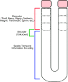
Fig. 6.. Modular division of steps for somite segmentation.
The spatiotemporal control of somite segmentation can be divided into three different modular steps: (i) information encoding by the clock and oscillatory gradient, (ii) decoding this information, and (iii) executing the segmentation decision.
Fig. 7.
Fig. 7.. Timeline of notable discoveries and model developments.
The timeline of somitogenesis, presenting models (top) and molecular discoveries (bottom). The models refer to preceding figure panels where applicable.

References

    1. Pourquie O., A brief history of the segmentation clock. Dev. Biol. 485, 24–36 (2022). - PubMed
    1. Piatkowska A. M., Evans S. E., Stern C. D., Cellular aspects of somite formation in vertebrates. Cells Dev. 168, 203732 (2021). - PubMed
    1. Gomez C., Ozbudak E. M., Wunderlich J., Baumann D., Lewis J., Pourquie O., Control of segment number in vertebrate embryos. Nature 454, 335–339 (2008). - PubMed
    1. Cooke J., Control of somite number during morphogenesis of a vertebrate, Xenopus laevis. Nature 254, 196–199 (1975). - PubMed
    1. Cooke J., Scale of body pattern adjusts to available cell number in amphibian embryos. Nature 290, 775–778 (1981). - PubMed

Publication types