. 2024 Feb 14;16(734):eadg7162.
doi: 10.1126/scitranslmed.adg7162. Epub 2024 Feb 14.

Mis-spliced transcripts generate de novo proteins in TDP-43-related ALS/FTD

Affiliations

Mis-spliced transcripts generate de novo proteins in TDP-43-related ALS/FTD

Sahba Seddighi et al. Sci Transl Med. .

Abstract

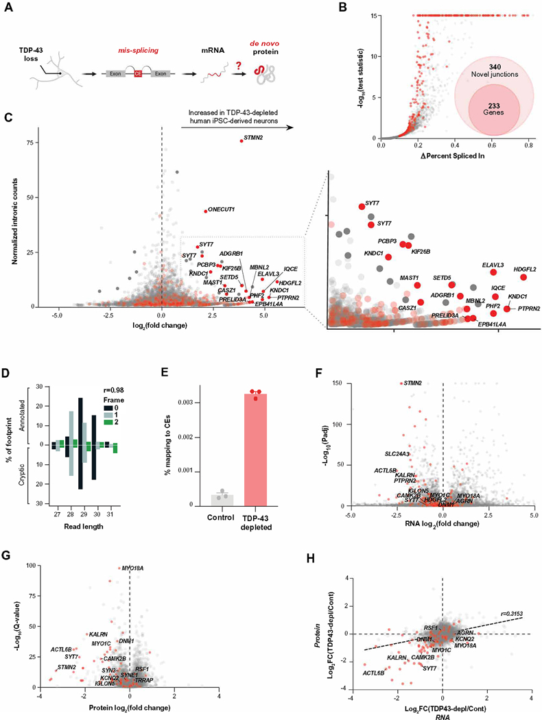
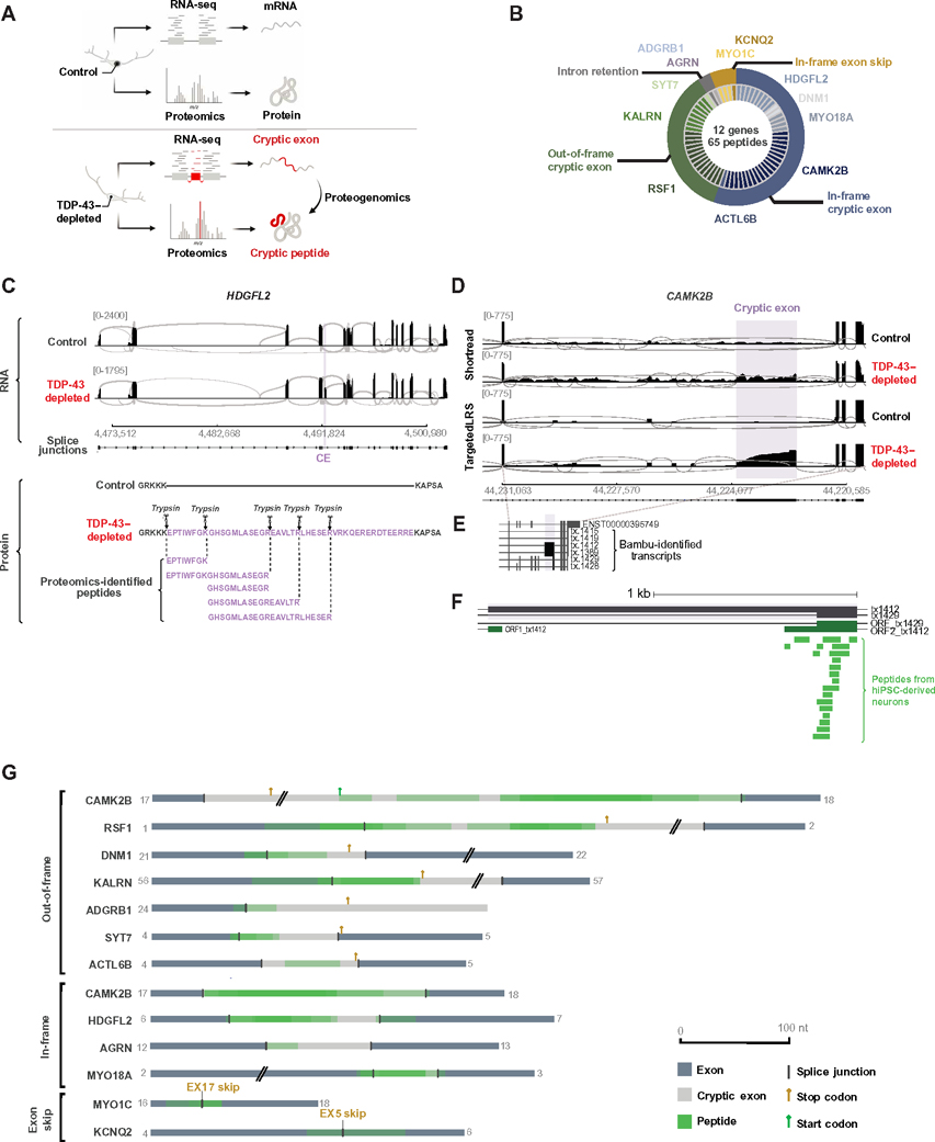
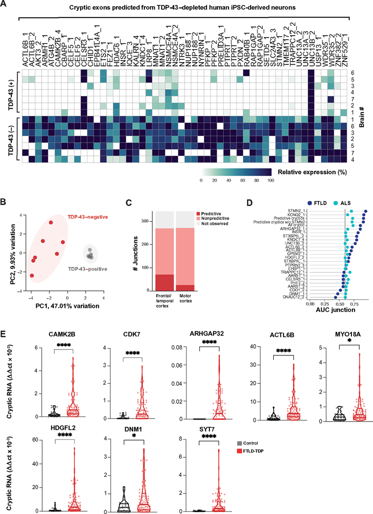
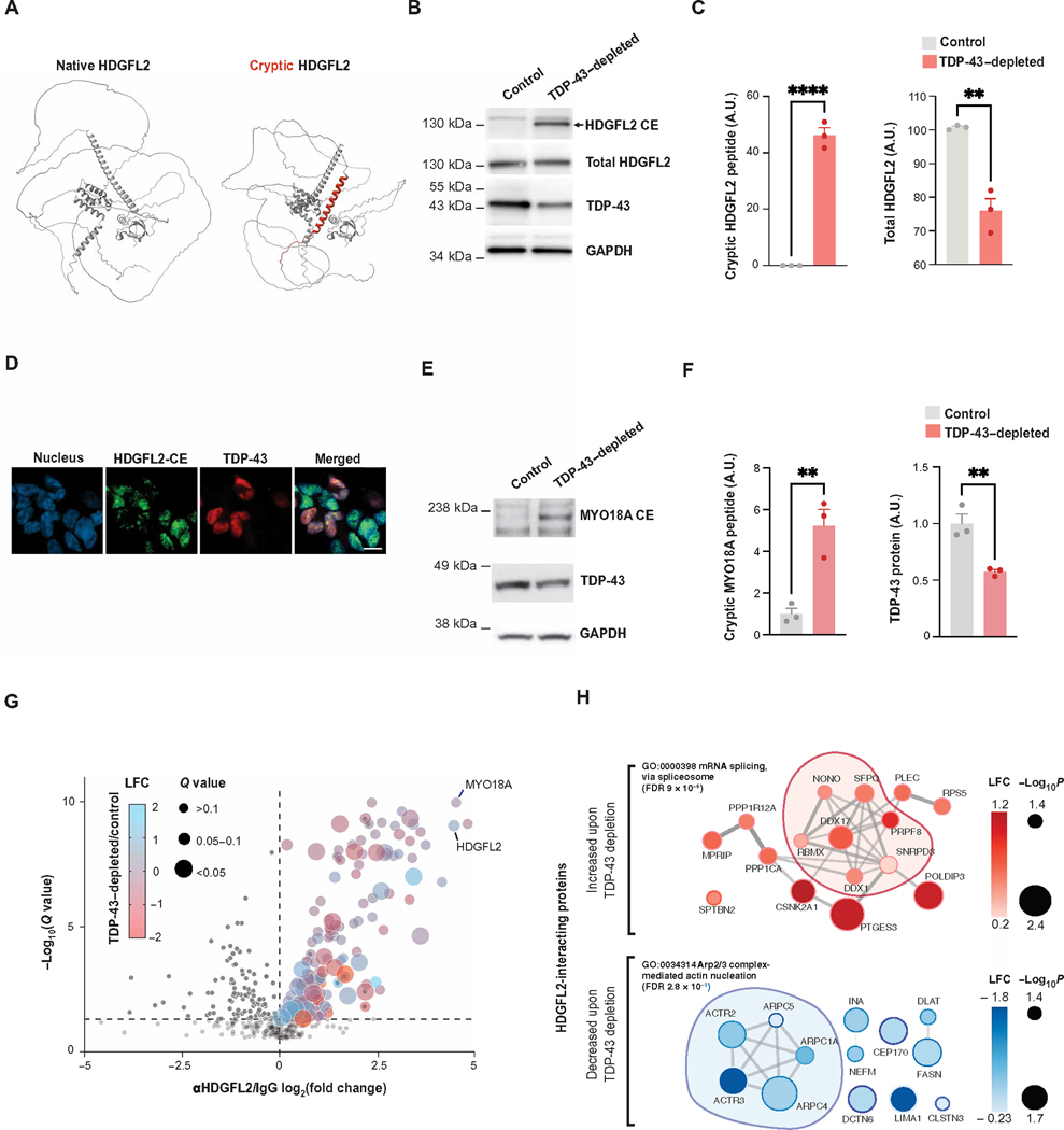
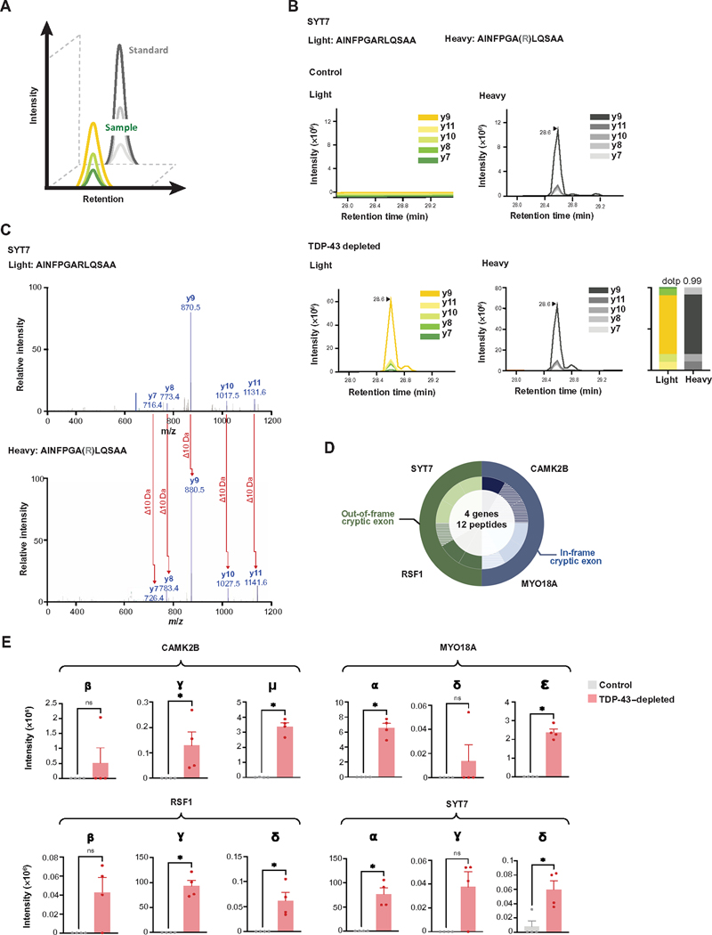
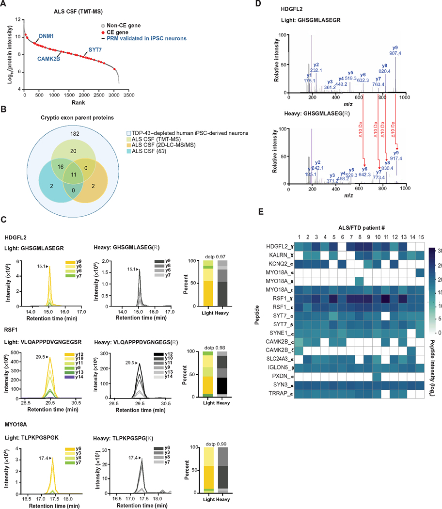
Functional loss of TDP-43, an RNA binding protein genetically and pathologically linked to amyotrophic lateral sclerosis (ALS) and frontotemporal dementia (FTD), leads to the inclusion of cryptic exons in hundreds of transcripts during disease. Cryptic exons can promote the degradation of affected transcripts, deleteriously altering cellular function through loss-of-function mechanisms. Here, we show that mRNA transcripts harboring cryptic exons generated de novo proteins in TDP-43-depleted human iPSC-derived neurons in vitro, and de novo peptides were found in cerebrospinal fluid (CSF) samples from patients with ALS or FTD. Using coordinated transcriptomic and proteomic studies of TDP-43-depleted human iPSC-derived neurons, we identified 65 peptides that mapped to 12 cryptic exons. Cryptic exons identified in TDP-43-depleted human iPSC-derived neurons were predictive of cryptic exons expressed in postmortem brain tissue from patients with TDP-43 proteinopathy. These cryptic exons produced transcript variants that generated de novo proteins. We found that the inclusion of cryptic peptide sequences in proteins altered their interactions with other proteins, thereby likely altering their function. Last, we showed that 18 de novo peptides across 13 genes were present in CSF samples from patients with ALS/FTD spectrum disorders. The demonstration of cryptic exon translation suggests new mechanisms for ALS/FTD pathophysiology downstream of TDP-43 dysfunction and may provide a potential strategy to assay TDP-43 function in patient CSF.


Conflict of interest statement

Competing interests: The participation of M.A.N., Z.L., and S.I.S. in this project was part of a competitive contract awarded to Data Tecnica International LLC by the NIH to support open science research. M.A.N. also currently serves as an advisor for Character Biosciences and Neuron23 Inc. N.P.S. consults for Emtherapro. B.O. serves as a consultant for Columbia University/Tsumura Inc., MediciNova, Biogen, UniQure, Amylyx, and Mitsubishi. L. Petrucelli consults for Expansion Therapeutics.

Figures

Fig. 1.
Fig. 1.. Transcriptional and proteomic analysis of TDP-43–depleted human iPSC–derived neurons.
(A) Overview of CE RNA and potential de novo protein production due to pathological mis-splicing in TDP-43–depleted human iPSC–derived neurons. (B) Shown is differential splicing in TDP-43–depleted human iPSC–derived neurons compared with human iPSC–derived control neurons expressing normal amounts of TDP-43. CE genes are shown in red. (C) Differential ribosome footprint density of intronic mRNA is shown for TDP-43–depleted human iPSC–derived neurons compared with human iPSC–derived control neurons. CE genes are shown in red. (D) Shown is the percentage abundance of each footprint type, defined by length and sub-codon position, for annotated coding sequences (positive Y axis) and a subset of high-confidence CEs, which are predicted to be translated (negative Y axis). Pearson’s correlation between the values for annotated and cryptic transcripts is shown. Data are from one TDP-43–depleted human iPSC–derived neuronal cell line. (E) Shown is the percentage of footprints aligning to the subset of CEs used in (C) for three TDP-43–depleted and three control human iPSC–derived neuronal samples. (F) Differential transcript abundance was quantified in TDP-43–depleted and control human iPSC–derived neurons using total short-read RNA-seq. CE genes are shown in red. (G) Differential protein abundance in TDP-43–depleted and control human iPSC–derived neurons was quantified using mass spectrometry proteomics. CE genes are shown in red. (H) Quantification of mRNA (RNA-seq) and protein by data-independent acquisition (DIA) proteomics of TDP-43–depleted and control human iPSC–derived neurons is shown. CEs are shown in red.
Fig. 2.
Fig. 2.. Formation of de novo cryptic peptides from mis-spliced transcripts in TDP-43–depleted human iPSC–derived neurons.
(A) Proteogenomic pipeline used to identify de novo cryptic peptides caused by RNA mis-splicing in TDP-43–depleted human iPSC–derived neurons. (B) Proteogenomic analysis of TDP-43–depleted human iPSC–derived neurons identified 65 putative trypsin-digested cryptic peptides across 12 genes. The outer circle of the pie chart represents genes. The inner circle represents the number of putative trypsin-digested cryptic peptides that were identified for each gene. (C) Representative sashimi plots showing the inclusion of an in-frame CE (purple) in HDGFL2 transcripts in TDP-43–depleted human iPSC–derived neurons. The amino acid sequence of the putative translation of this CE is shown below the sashimi plot, with trypsin cleavage sites and proteogenomic-identified cryptic peptides annotated. (D) Representative sashimi plots comparing Illumina short-read RNA-seq versus Nanopore long-read sequencing of the CAMK2B transcript in control and TDP-43–depleted human iPSC–derived neurons. The CE expressed in TDP-43–depleted human iPSC–derived neurons is highlighted in purple. (E) Precise mapping of CAMK2B exon junctions and splice isoforms of transcripts in TDP-43–depleted human iPSC–derived neurons using Nanopore long-read sequencing. (F) Tryptic-cryptic peptides (green) identified using the proteogenomic pipeline are mapped to an in-frame CE in CAMK2B identified using Nanopore long-read sequencing. (G) Graphical representation of proteogenomic-identified cryptic peptides mapped to transcripts from Nanopore long-read sequencing. Two genes, MYO1C and KCNQ2, contained an in-frame exon skipping event upon TDP-43 loss. Four genes, HDGFL2, AGRN, MYO18A, and CAMK2B, contained in-frame CEs. An out-of-frame CAMK2B CE was also identified. Canonical upstream and downstream exons are in dark gray. CEs are in light gray. Cryptic peptide locations are overlaid in green.
Fig. 3.
Fig. 3.. TDP-43 cryptic exons in TDP-43–depleted human iPSC–derived neurons predict TDP-43 pathology in postmortem brain tissue.
(A) Heatmap showing the relative abundance of CEs predicted from TDP-43–depleted human iPSC–derived neurons in FACS-sorted TDP-43–positive and TDP-43–negative neuronal nuclei preparations from postmortem ALS/FTD cortical samples (n = 7) (17). The “percent spliced in” (PSI) value represents the ratio of transcripts that include a splicing event versus the total number of transcripts. We defined enriched junctions as those with PSI in TDP-43–negative nuclei greater than twice the PSI in TDP-43–positive nuclei (mean TDP-43–negative PSI > 0.10). The top 50 most expressed cryptic splice junctions in TDP-43–negative nuclei, compared with TDP-43–positive nuclei, are shown, and cases were organized by unsupervised hierarchical clustering based on CE PSI. (B) Principal components analysis (PCA) on PSI of 230 predicted TDP-43 CEs in TDP-43–positive and TDP-43–negative neuronal nuclei from ALS/FTD postmortem cortical samples. (C) Bar plot showing the number of predicted CEs detected in bulk RNA-seq of ALS/FTD postmortem brain tissue samples from the NYGC biobank. CEs were classified by whether they were observed in postmortem brain tissue and were detected/nonpredictive or detected/predictive of TDP-43 pathology versus non–TDP-43 pathology. (D) AUC is shown for predicted CEs that identified TDP-43 pathology in FTLD frontal/temporal cortex and ALS motor cortex postmortem tissue from patients with ALS/FTLD patients. Meta-expression scores for all CEs, only predictive exons (AUC > = 0.6), and predictive exons excluding STMN2 expression are shown. (E) Shown is quantitative RT-PCR–based validation of eight CEs in an independent set of postmortem frontal cortex brain samples from patients with FTLD-TDP (n = 89) and age-matched controls with non-neurological disease (n = 27). Data are presented as means ± SEM. P values are from the Mann-Whitney test: *P < 0.05 and ****P ≤ 0.0001.
Fig. 4.
Fig. 4.. Cryptic peptides in TDP-43–depleted human iPSC–derived neurons alter the protein interactome.
(A) Predicted structure of HDGFL2 (using AlphaFold), annotated with the predicted cryptic peptide induced by TDP-43 depletion highlighted in red. (B) Antibody-based detection of an HDGFL2 cryptic peptide in TDP-43–depleted human iPSC–derived neurons. Representative Western blot shows a band of the expected molecular weight of HDGFL2-CE specifically in TDP-43–depleted but not control human iPSC–derived neurons. (C) Quantification of HDGFL2-CE and total HDGFL2 in Western blot from (B) (n = 3, two-sample t test, **P < 0.01 and ****P < 0.0001; Shapiro-Wilk test for normality, P > 0.05 not significant). (D) Immunofluorescence staining highlights the selective expression of the HDGFL2 cryptic peptide in TDP-43–depleted human iPSC–derived neurons (scale bar,10 μm). (E) Antibody-based detection of a MYO18A cryptic peptide in TDP-43–depleted human iPSC–derived neurons. Representative Western blot shows a band of the expected molecular weight of the MYO18A-CE specifically in TDP-43–depleted human iPSC–derived neurons. (F) Quantification of MYO18A-CE and TDP-43 protein expression (n = 3, two-sample t test, **P < 0.01; Shapiro-Wilk test for normality, P > 0.05 not significant). (G) Affinity purification mass spectrometry analysis of HDGFL2 protein-protein interactions in TDP-43–depleted and control human iPSC–derived neurons. A volcano plot of coimmunoprecipitated proteins using anti-HDGFL2 antibody versus control IgG is shown. The dot color reflects log fold change (LFC) in TDP-43–depleted versus control human iPSC–derived neurons, and the dot size reflects the adjusted P value (q value). (H) STRING diagram of proteins whose interactions with HDGFL2 were significantly altered by TDP-43–depleted versus control human iPSC–derived neurons (Padj < 0.05). The dot color reflects LFC in TDP-43–depleted versus control human iPSC–derived neurons.
Fig. 5.
Fig. 5.. Scalable cryptic peptide validation in TDP-43–depleted human iPSC–derived neurons by targeted proteomics.
(A) Schematic of PRM-MS. Co-elution of SIL peptides allows for sensitive measurement of corresponding endogenous peptides. (B) PRM-MS assay using a synthetic SIL peptide internal standard identifies a cryptic peptide in SYT7 in TDP-43–depleted but not in control human iPSC–derived neurons. The spectral plot of heavy standards and light (endogenous) y ions from an SYT7 cryptic peptide is shown, with accompanying dot product (dotp), which indicates the correlation between the peptide fragment-ion peak areas and theoretical spectra. (C) Corresponding mass spectra of endogenous and heavy peptide standards of the SYT7 cryptic peptide in TDP-43–depleted human iPSC–derived neurons. (D) The detection of 12 trypsin-digested cryptic peptides across four genes using single-shot PRM assays in TDP-43–depleted human iPSC–derived neuronal lysates is shown. The outer circle represents the gene, and the inner circle represents the number of cryptic peptides detected by PRM per gene. Hatched color signifies the successful detection of one to two y ions; solid color signifies the detection of three or more y ions. (E) Quantification of cryptic peptide expression in TDP-43–depleted and control human iPSC–derived neurons using PRM assays. n = 4 replicates per sample. Mann-Whitney U test. *P < 0.05, ns, not significant.
Fig. 6.
Fig. 6.. Cryptic peptides are present in CSF samples from patients with ALS.
(A) Rank plot of proteins detected in CSF samples from 24 patients with ALS. Forty-seven CE genes that were predicted in TDP-43–depleted human iPSC–derived neurons are shown in red. PRM-validated cryptic peptides expressing genes (from human iPSC–derived neuron studies) are annotated in blue text. (B) Euler diagram of three CSF proteomics datasets versus CEs predicted in TDP-43–depleted human iPSC–derived neurons. (C) Representative spectra of three heavy (standard) and light (endogenous) cryptic peptides detected in CSF from patients with ALS/FTD spectrum disorders (n = 13 ALS, n = 1 ALS/FTD, n = 1 ALS/mild cognitive impairment). Also shown is the dotp, which indicates the correlation between the peptide fragment-ion peak areas and theoretical spectra. The spectra for 15 additional cryptic peptides in ALS/FTD CSF samples are shown in fig. S9A. (D) MS/MS spectrum of a cryptic peptide in HDGFL2 that corresponds to the reference peptide (top) and endogenous peptide (bottom) detected in ALS/FTD CSF samples. (E) Heatmap of AUC intensities of 18 cryptic peptides from 13 different proteins in CSF samples from 15 patients with ALS/FTD.

Update of

  • Mis-spliced transcripts generate de novo proteins in TDP-43-related ALS/FTD.
    Seddighi S, Qi YA, Brown AL, Wilkins OG, Bereda C, Belair C, Zhang Y, Prudencio M, Keuss MJ, Khandeshi A, Pickles S, Hill SE, Hawrot J, Ramos DM, Yuan H, Roberts J, Kelmer Sacramento E, Shah SI, Nalls MA, Colon-Mercado J, Reyes JF, Ryan VH, Nelson MP, Cook C, Li Z, Screven L, Kwan JY, Shantaraman A, Ping L, Koike Y, Oskarsson B, Staff N, Duong DM, Ahmed A, Secrier M, Ule J, Jacobson S, Rohrer J, Malaspina A, Glass JD, Ori A, Seyfried NT, Maragkakis M, Petrucelli L, Fratta P, Ward ME. Seddighi S, et al. bioRxiv [Preprint]. 2023 Jan 23:2023.01.23.525149. doi: 10.1101/2023.01.23.525149. bioRxiv. 2023. Update in: Sci Transl Med. 2024 Feb 14;16(734):eadg7162. doi: 10.1126/scitranslmed.adg7162. PMID: 36747793 Free PMC article. Updated. Preprint.

References

    1. Neumann M, Tolnay M, Mackenzie IRA, The molecular basis of frontotemporal dementia. Expert Rev. Mol. Med. 11, e23 (2009). - PubMed
    1. Meneses A, Koga S, O’Leary J, Dickson DW, Bu G, Zhao N, TDP-43 pathology in Alzheimer’s Disease. Neurodegeneration 16, 84 (2021). - PMC - PubMed
    1. Van Deerlin VM, Leverenz JB, Bekris LM, Bird TD, Yuan W, Elman LB, Clay D, Wood EMC, Chen-Plotkin AS, Martinez-Lage M, Steinbart E, McCluskey L, Grossman M, Neumann M, Wu IL, Yang WS, Kalb R, Galasko DR, Montine TJ, Trojanowski JQ, Lee VMY, Schellenberg GD, Yu CE, TARDBP mutations in amyotrophic lateral sclerosis with TDP-43 neuropathology: A genetic and histopathological analysis. Lancet Neurol. 7, 409–416 (2008). - PMC - PubMed
    1. Borroni B, Bonvicini C, Alberici A, Buratti E, Agosti C, Archetti S, Papetti A, Stuani C, Di Luca M, Gennarelli M, Padovani A, Mutation withinTARDBPleads to frontotemporal dementia without motor neuron disease. Hum. Mutat. 30, E974–E983 (2009). - PubMed
    1. Halliday G, Bigio EH, Cairns NJ, Neumann M, MacKenzie IRA, Mann DMA, Mechanisms of disease in frontotemporal lobar degeneration: Gain of function versus loss of function effects. Acta Neuropathol. 124, 373–382 (2012). - PMC - PubMed

Publication types