. 2024 Feb 16;19(2):516-525.
doi: 10.1021/acschembio.3c00686. Epub 2024 Jan 26.

Expanding the Genetic Code of Xenopus laevis Embryos

Affiliations

Expanding the Genetic Code of Xenopus laevis Embryos

Wes Brown et al. ACS Chem Biol. .

Abstract

The incorporation of unnatural amino acids into proteins through genetic code expansion has been successfully adapted to African claw-toed frog embryos. Six unique unnatural amino acids are incorporated site-specifically into proteins and demonstrate robust and reliable protein expression. Of these amino acids, several are caged analogues that can be used to establish conditional control over enzymatic activity. Using light or small molecule triggers, we exhibit activation and tunability of protein functions in live embryos. This approach was then applied to optical control over the activity of a RASopathy mutant of NRAS, taking advantage of generating explant cultures from Xenopus. Taken together, genetic code expansion is a robust approach in the Xenopus model to incorporate novel chemical functionalities into proteins of interest to study their function and role in a complex biological setting.


Conflict of interest statement

The authors declare no competing financial interest.

Figures
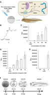
Figure 1
Figure 1
Genetic code expansion in Xenopus. (A) Injection and UAA incorporation into a protein of interest (POI) in embryos. (B) Structure of 1. (C) Titration of 1 in the injection solution and incorporation into Rluc95UAG. (D) Incorporation of 1 (10 mM) over the first 72 h of development. (E) Incorporation of 1 into Rluc95UAG at 16 °C. All bars are means, and error bars are standard deviations from three embryos. (F) Nieuwkoop Faber (NF) stages of Xenopus embryos at certain time points at 23 °C. Panel F reproduced with permission from ref (37). Copyright 2022 The Company of Biologists. NT = nontreated embryo control. hpf = hours post-fertilization.
Figure 2
Figure 2
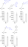
Genetic encoding of UAAs 26 in Xenopus embryos. Structures of 26. Blue represents caging groups that are removed after treatment with 405 nm light or a small molecule trigger. Incorporation of 26 into the luciferase reporter was accomplished after injection of all components, including 2, 4, and 5, while UAAs 3 and 6 were added to the media. Embryos were incubated at 23 °C, and assays were conducted at 24 hpf. Bars represent means, and error bars represent standard deviations of measurements from three independent embryos.
Figure 3
Figure 3
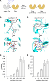
Conditional control of enzymatic activity with photocaged UAAs 2 and 3. (A) Luciferase enzymatic activation in Xenopus embryos with light. Structural representation of caging of the Fluc active site with either (B) 2 or (D) 3 before and after irradiation (PDB: 4G36). Incorporation of (C) 2 or (E) 3 into the dual luciferase reporter and irradiation for increasing duration with a 405 nm light at 24 hpf. Bars represent means, and error bars represent standard deviations of measurements from three independent embryos.
Figure 4
Figure 4
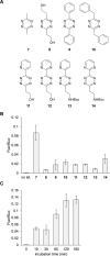
Conditional control of enzymatic activity through the incorporation of 4. (A) Chemical structures of the different tetrazines tested for decaging of 4in vivo. (B) Tetrazine screen of dual-luciferase reporter activation. (C) Tetrazine 7 incubation timecourse. Bars represent mean, and error bars represent standard deviation of measurements from three independent embryos. No tet = no tetrazine control.
Figure 5
Figure 5
Conditional control of enzymatic activity through phosphine-based decaging of 5 incorporated into protein. (A) Chemical structures of the different phosphines tested. (B) Phosphine screen of dual-luciferase reporter activation. (C) Incubation timecourse with 16. Bars represent mean, and error bars represent standard deviation of measurements from three independent embryos. No phos. = no phosphine control.
Figure 6
Figure 6
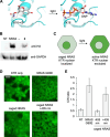
Optical control of NRAS activity using photocaged lysine 2. (A) Structural representation of caging of the NRAS active site with 2 (PDB: 5UHV). (B) Western blot of incorporation of 2 into NRAS. (C) Caging of NRAS blocks its activity in the absence of light, and the ERKKTR-Clover reporter is nuclear localized. Irradiation at 405 nm activates NRAS, which leads to ERK activation and nuclear exclusion of the KTR. (D) Representative confocal images of the KTR reporter for each condition. Scale bar: 20 μm. (E) Quantification of the fluorescent cytosol/nucleus ratio. Bars represent means, and error bars represent standard deviations of measurements from 15 cells and three different embryos.

Similar articles

Cited by

References

    1. Chung C. Z.; Amikura K.; Söll D. Using Genetic Code Expansion for Protein Biochemical Studies. Frontiers in Bioengineering and Biotechnology 2020, 8, 59857710.3389/fbioe.2020.598577. - DOI - PMC - PubMed
    1. Courtney T.; Deiters A. Recent advances in the optical control of protein function through genetic code expansion. Curr. Opin Chem. Biol. 2018, 46, 99–107. 10.1016/j.cbpa.2018.07.011. - DOI - PMC - PubMed
    1. Schmidt M.; Summerer D. Genetic code expansion as a tool to study regulatory processes of transcription. Frontiers in Chemistry 2014, 2, 7.10.3389/fchem.2014.00007. - DOI - PMC - PubMed
    1. Shandell M. A.; Tan Z.; Cornish V. W. Genetic Code Expansion: A Brief History and Perspective. Biochemistry 2021, 60 (46), 3455–3469. 10.1021/acs.biochem.1c00286. - DOI - PMC - PubMed
    1. Courtney T. M.; Deiters A. Optical control of protein phosphatase function. Nat. Commun. 2019, 10 (1), 4384.10.1038/s41467-019-12260-z. - DOI - PMC - PubMed

Publication types