. 2024 Feb 13;19(2):224-238.
doi: 10.1016/j.stemcr.2023.12.011. Epub 2024 Jan 25.

Proinflammatory phenotype of iPS cell-derived JAK2 V617F megakaryocytes induces fibrosis in 3D in vitro bone marrow niche

Affiliations

Proinflammatory phenotype of iPS cell-derived JAK2 V617F megakaryocytes induces fibrosis in 3D in vitro bone marrow niche

Niclas Flosdorf et al. Stem Cell Reports. .

Abstract

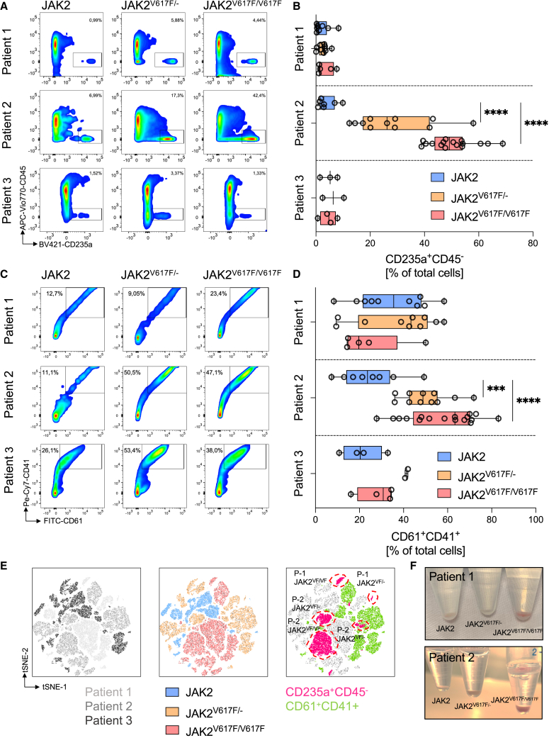
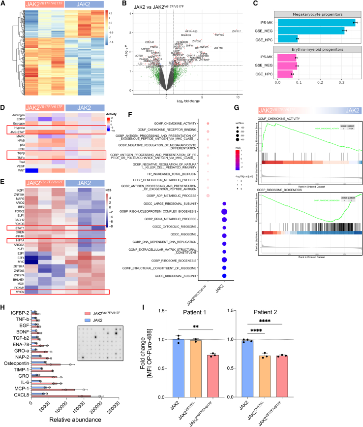
The myeloproliferative disease polycythemia vera (PV) driven by the JAK2 V617F mutation can transform into myelofibrosis (post-PV-MF). It remains an open question how JAK2 V617F in hematopoietic stem cells induces MF. Megakaryocytes are major players in murine PV models but are difficult to study in the human setting. We generated induced pluripotent stem cells (iPSCs) from JAK2 V617F PV patients and differentiated them into megakaryocytes. In differentiation assays, JAK2 V617F iPSCs recapitulated the pathognomonic skewed megakaryocytic and erythroid differentiation. JAK2 V617F iPSCs had a TPO-independent and increased propensity to differentiate into megakaryocytes. RNA sequencing of JAK2 V617F iPSC-derived megakaryocytes reflected a proinflammatory, profibrotic phenotype and decreased ribosome biogenesis. In three-dimensional (3D) coculture, JAK2 V617F megakaryocytes induced a profibrotic phenotype through direct cell contact, which was reversed by the JAK2 inhibitor ruxolitinib. The 3D coculture system opens the perspective for further disease modeling and drug discovery.


Conflict of interest statement

Declaration of interests W.W. and V.T. are involved in Cygenia GmbH (www.cygenia.com), which provides services for DNA methylation analysis to other scientists. S.K. reports funding from Novartis, Bristol-Myers Squibb, and Janssen/Geron; advisory board honoraria from Pfizer, Incyte, Ariad, Novartis, AOP Pharma, BMS, Celgene, Geron, Janssen, CTI, Roche, Baxalta, GSK, Sierra Oncology, and Sanofi; a patent for Bromodomain and Extra-Terminal (BET) inhibitor at RWTH Aachen University; honoraria from Novartis, Bristol Myers Squibb, Celgene, Geron, Janssen, Pfizer, Incyte, Ariad, Shire, Roche, and AOP Pharma; and other financial support (e.g., travel support) from Alexion, Novartis, Bristol Myers Squibb, Incyte, Ariad, AOP Pharma, Baxalta, CTI, Pfizer, Sanofi, Celgene, Shire, Janssen, Geron, Abbvie, Imago Biosciences, Sierra Oncology, GSK, and Karthos. T.H.B. served as a consultant for Janssen, Merck, Novartis, and Pfizer; received research funding form Novartis and Pfizer; and received honoraria from Janssen, Merck, Novartis, and Pfizer.

Figures

None
Graphical abstract
Figure 1
Figure 1
Patient-derived JAK2V617F/− and JAK2V617F/V617F iPSC differentiation into MKs (A) Representative allele-specific PCR analysis and Sanger sequencing of iPSCs without JAK2 mutation and monoallelic and biallelic JAK2 V617F mutation (JAK2, JAK2V617F/−, and JAK2V617F/V617F, respectively). The G>T transition in JAK2 V617F mutated clones is indicated. W, water control. (B) iPSC differentiation into EBs, hematopoietic progenitors, and MKs by specific cytokines as indicated. (C) JAK2 V617F mutation increased the total cell numbers produced per EB (day 14) of all 3 patients. Each dot represents an independent experiment (n = 11–12). (D) The iPSC-MKs showed MK morphology with budding and proplatelet formation (Romanowski staining, patient 2). Scale bars, 50 μm. (E) Images of iPSC-MKs by TEM (patient 2). DMS, demarcation membrane system; N, nucleus. Asterisks indicate proplatelet protrusions. White arrows mark granules. Scale bars, 1,000 nm. See also Figures S1–S3.
Figure 2
Figure 2
Hematopoietic differentiation of JAK2V617F/− and JAK2V617F/V617F iPSCs recapitulates skewed myeloid differentiation of PV patients (A and B) Increased erythropoiesis of patient 2 iPSCs with JAK2 V617F mutation in MK differentiation (day 14) independent of EPO by increased frequency of CD235a+CD45 cells in flow cytometry. (C and D) Production of CD61+CD41+ MKs for all iPSC clones of all of the patients, which was particularly prominent for the JAK2 V617F mutated cells of patient 2. (E) tSNE analysis of events from all 3 patients revealed more prominent clusters of CD235a+CD45 cells overlapping especially with the JAK2V617F/− and JAK2V617F/V617F cells (patients 1 and 2). (F) Increased erythropoiesis in JAK2V617F/V617F cells as evidenced by red cell pellet at day 14 of differentiation as in (A)–(E). (B and D) Each dot represents an independent experiment (patient 1, n = 5–10; patient 2, n = 7–17; patient 3, n = 2–4). See also Figure S4.
Figure 3
Figure 3
JAK2V617F/− and JAK2V617F/V617F iPSCs develop into MKs independently of TPO (A) Flow cytometry analysis (CD41, CD42b, CD61) of JAK2 V617F MKs of patient 2 in response to TPO (2 days) on day 14 of MK differentiation shows independence of TPO in JAK2V617F/V617F cells. (B) Quantification of CD61+CD41+ and CD41+CD42b+ cells in response of TPO as in (A) of patients 1–3. JAK2V617F/V617F depicts data of patient 2. JAK2 V617F cells show TPO independent increased production of MKs, whereas MK production of unmutated cells was TPO dependent. Each dot represents an independent experiment (JAK2, n = 13–15; JAK2V617F/−, n = 10–12; JAK2V617F/V617F, n = 5–11).
Figure 4
Figure 4
JAK2V617F/V617F iPS MKs express an inflammatory gene signature iPSCs with homozygous JAK2 V617F mutation and without mutation were differentiated into MKs (day 14) and subjected to bulk RNA sequencing analysis (N = 3, patient 2). (A and B) JAK2V617F/V617F MKs showed 279 significantly differentially expressed genes (187 downregulated and 92 upregulated genes, respectively) and are represented in heatmap and volcano plot format (A and B, respectively). (C) CIBERSORT analysis identified iPSC-MKs of all genotypes as MK and erythromyeloid progenitors (GSE_Meg and GSE_HPC refer to MK and hematopoietic progenitor [HPC] datasets). (D) PROGENy analysis revealed prominent upregulation of the JAK-STAT and also upregulation of hypoxia, TNF-⍺, and TGF-β pathways (red boxes). (E) DoRothEA analysis showed downregulation of TFs MYCN and upregulation STAT1 and HIF1A (red boxes). (F and G) GSEA analysis showed, among others, the positive enrichment of chemokines and the negative enrichment of ribosome-associated genes. (H) ELISA of multiple chemokines shows prominent upregulation of inflammatory cytokines in JAK2V617F/V617F MKs (CXCL8/IL-8, MCP-1, IL-6). (I) Uptake of OP-Puro in nascent peptide chains was decreased in JAK2V617F/V617F MKs (patients 1 and 2; n = 2–3 and n = 3–4, respectively). See also Figure S5.
Figure 5
Figure 5
JAK2V617F/− and JAK2V617F/V617F MKs induce profibrotic gene expression in stromal cells in a 3D coculture model (A) Schematic representation of coculturing MSCs and MKs on 3D scaffolds. (B) Representative scanning electron microscopy image of MEW printed fiber scaffolds with respective scaffold characteristics. (C) MSC growing on the scaffolds (left) and in coculture (right). JAK2 V617F MKs (CD42b, arrows) were in close proximity to MSC (GFP). (D) ACTA2, FAP, GLI1, COL1A1, and TGF-β1 gene expression in MSC was upregulated in coculture with JAK2 V617F MKs and CXCL12 gene expression was downregulated (patients 1 and 2). Ruxolitinib treatment abolished the activity of JAK2V617F/V617F MKs. Each dot represents an independent experiment (JAK2, n = 4–10; JAK2V617F/−, n = 6–9; JAK2V617F/V617F, n = 3–9; ruxolitinib, n = 2–4). See also Figure S6.

References

    1. Ackermann M., Liebhaber S., Klusmann J.H., Lachmann N. Lost in translation: pluripotent stem cell-derived hematopoiesis. EMBO Mol. Med. 2015;7:1388–1402. - PMC - PubMed
    1. Alsinet C., Primo M.N., Lorenzi V., Bello E., Kelava I., Jones C.P., Vilarrasa-Blasi R., Sancho-Serra C., Knights A.J., Park J.E., et al. Robust temporal map of human in vitro myelopoiesis using single-cell genomics. Nat. Commun. 2022;13:2885. - PMC - PubMed
    1. Arber D.A., Orazi A., Hasserjian R., Thiele J., Borowitz M.J., Le Beau M.M., Bloomfield C.D., Cazzola M., Vardiman J.W. The 2016 revision to the World Health Organization classification of myeloid neoplasms and acute leukemia. Blood. 2016;127:2391–2405. - PubMed
    1. Balduini A., Badalucco S., Pugliano M.T., Baev D., De Silvestri A., Cattaneo M., Rosti V., Barosi G. In vitro megakaryocyte differentiation and proplatelet formation in Ph-negative classical myeloproliferative neoplasms: distinct patterns in the different clinical phenotypes. PLoS One. 2011;6:e21015. - PMC - PubMed
    1. Baumeister J., Chatain N., Hubrich A., Maié T., Costa I.G., Denecke B., Han L., Küstermann C., Sontag S., Seré K., et al. Hypoxia-inducible factor 1 (HIF-1) is a new therapeutic target in JAK2V617F-positive myeloproliferative neoplasms. Leukemia. 2020;34:1062–1074. - PubMed

Publication types