. 2024 Feb;5(2):e109-e118.
doi: 10.1016/S2666-5247(23)00259-8. Epub 2024 Jan 24.

2020 Ebola virus disease outbreak in Équateur Province, Democratic Republic of the Congo: a retrospective genomic characterisation

Affiliations

2020 Ebola virus disease outbreak in Équateur Province, Democratic Republic of the Congo: a retrospective genomic characterisation

Eddy Kinganda-Lusamaki et al. Lancet Microbe. 2024 Feb.

Abstract

Background: The Democratic Republic of the Congo has had 15 Ebola virus disease (EVD) outbreaks, from 1976 to 2023. On June 1, 2020, the Democratic Republic of the Congo declared an outbreak of EVD in the western Équateur Province (11th outbreak), proximal to the 2018 Tumba and Bikoro outbreak and concurrent with an outbreak in the eastern Nord Kivu Province. In this Article, we assessed whether the 11th outbreak was genetically related to previous or concurrent EVD outbreaks and connected available epidemiological and genetic data to identify sources of possible zoonotic spillover, uncover additional unreported cases of nosocomial transmission, and provide a deeper investigation into the 11th outbreak.

Methods: We analysed epidemiological factors from the 11th EVD outbreak to identify patient characteristics, epidemiological links, and transmission modes to explore virus spread through space, time, and age groups in the Équateur Province, Democratic Republic of the Congo. Trained field investigators and health professionals recorded data on suspected, probable, and confirmed cases, including demographic characteristics, possible exposures, symptom onset and signs and symptoms, and potentially exposed contacts. We used blood samples from individuals who were live suspected cases and oral swabs from individuals who were deceased to diagnose EVD. We applied whole-genome sequencing of 87 available Ebola virus genomes (from 130 individuals with EVD between May 19 and Sept 16, 2020), phylogenetic divergence versus time, and Bayesian reconstruction of phylogenetic trees to calculate viral substitution rates and study viral evolution. We linked the available epidemiological and genetic datasets to conduct a genomic and epidemiological study of the 11th EVD outbreak.

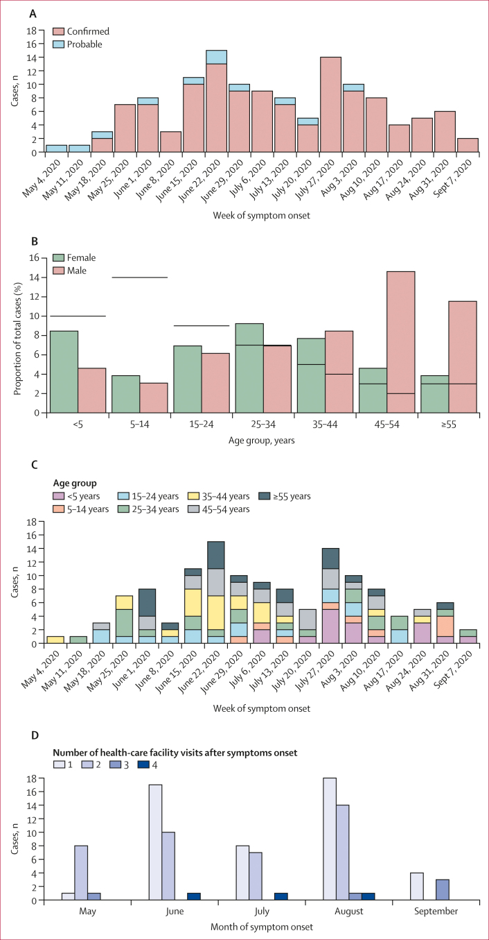
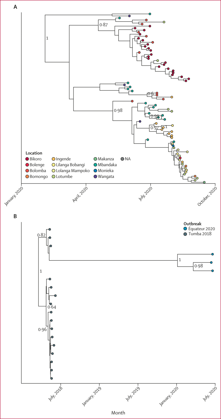
Findings: Between May 19 and Sept 16, 2020, 130 EVD (119 confirmed and 11 probable) cases were reported across 13 Équateur Province health zones. The individual identified as the index case reported frequent consumption of bat meat, suggesting the outbreak started due to zoonotic spillover. Sequencing revealed two circulating Ebola virus variants associated with this outbreak-a Mbandaka variant associated with the majority (97%) of cases and a Tumba-like variant with similarity to the ninth EVD outbreak in 2018. The Tumba-like variant exhibited a reduced substitution rate, suggesting transmission from a previous survivor of EVD.

Interpretation: Integrating genetic and epidemiological data allowed for investigative fact-checking and verified patient-reported sources of possible zoonotic spillover. These results demonstrate that rapid genetic sequencing combined with epidemiological data can inform responders of the mechanisms of viral spread, uncover novel transmission modes, and provide a deeper understanding of the outbreak, which is ultimately needed for infection prevention and control during outbreaks.

Funding: WHO and US Centers for Disease Control and Prevention.


Conflict of interest statement

Declaration of interests We declare no competing interests.

Figures

Figure 1
Figure 1
Ebola virus outbreaks in the Democratic Republic of the Congo, 1976–2022 (A) The distribution of Ebola virus disease outbreaks in the Democratic Republic of the Congo. Coloured circles identify locations of previous outbreaks and the size of circles represents the number of positive human cases. The map shows the affected 2020 health zones (orange and purple shading) and sites of the 2018 Équateur outbreak (orange shading). The red star indicates the location of the Kinshasa diagnostic laboratory during the Équateur Province 2020 outbreak. (B) The locations and prevalence of Ebola virus disease cases during the 2020 Ebola virus disease outbreak in the Équateur Province. The red stars indicate the location of the diagnostic and field laboratories during the Équateur Province 2020 outbreak (Mbandaka, Ingende, Itipo, Bikoro, and Bolomba).
Figure 2
Figure 2
Demographics of EVD cases during the 2020 Équateur Province outbreak (A) Epidemiological curve of confirmed and probable EVD cases over time. (B) Age distribution of confirmed and probable EVD cases by gender (the black horizontal bars represent the 2020 Democratic Republic of the Congo known age and gender population distribution from WHO). (C) Temporal age distribution of individuals with EVD. (D) Distribution of patients with EVD who visited multiple health-care facilities after symptoms onset. EVD=Ebola virus disease.
Figure 3
Figure 3
Social networks using interviews and contact tracing from the Équateur Province outbreak Each circle represents a single individual and is located at the date of symptom onset. Individuals from which genetic data are available are indicated with a box around the individuals’ identification number. Grey lines connecting nodes indicate social connections gathered from contact tracing interviews. The red arrow highlights the contact tracing chain associated with 2018 Tumba-like sequences. Grey arrows highlight examples of nosocomial transmission.
Figure 4
Figure 4
Ebola virus variants circulating during the 2020 Équateur Province outbreak (A) Inferred phylogenetic history using all available Mbandaka variant sequences. Posterior probabilities greater than 0·7 are labelled at internal nodes. (B) Inferred phylogenetic history of Tumba variant sequences. Posterior probabilities greater than 0·7 are labelled at internal nodes. (C) The genetic divergence from root versus collection date for the 2018 Équateur (Tumba variant) outbreak, including Tumba-like sequences from the 2020 Équateur Province EVD outbreak. The dashed line indicates the substitution rate for the 2018 Équateur outbreak and the dotted lines are 95% prediction intervals using only the 2018 Tumba sequences. EVD=Ebola virus disease. NA=not available.
Figure 4
Figure 4
Ebola virus variants circulating during the 2020 Équateur Province outbreak (A) Inferred phylogenetic history using all available Mbandaka variant sequences. Posterior probabilities greater than 0·7 are labelled at internal nodes. (B) Inferred phylogenetic history of Tumba variant sequences. Posterior probabilities greater than 0·7 are labelled at internal nodes. (C) The genetic divergence from root versus collection date for the 2018 Équateur (Tumba variant) outbreak, including Tumba-like sequences from the 2020 Équateur Province EVD outbreak. The dashed line indicates the substitution rate for the 2018 Équateur outbreak and the dotted lines are 95% prediction intervals using only the 2018 Tumba sequences. EVD=Ebola virus disease. NA=not available.
Figure 5
Figure 5
Epidemiological and phylogenetic networks showing details from the 2020 Équateur Province outbreak (A) Genetic data and contact tracing chains for co-circulating Mbandaka and Tumba EBOV variants. Filled circles represent single individuals and are located at symptom onset dates. Colours indicate residence location. The red box indicates a contact tracing chain containing Tumba-like EBOV sequences. Identification numbers with boxes around them show that genetic data were available, with shading in boxes indicating the genetic distance from the earliest sequence in the outbreak (with green showing little genetic distance and red showing greater genetic distance). (B) An example of nosocomial transmission from overlapping health-care facility stays (red arrow). Individual rdceqt003004 visited health-care facility B, concurrently with individual rdceqt000227 (who had EVD), before becoming positive for EVD. Filled circles represent single individuals and are located at symptom onset dates. Circle colours indicate residence location. Yellow bars represent length of stay at health-care facilities after EVD symptom onset. (C) Contact tracing data re-evaluated by combining genetic and epidemiological data. Metadata showing the identification of overlapping hospital stays at facility G between the individual rdceqt000001 (EVD case) and individual rdceqt000008 (red arrow). Individual rdceqt000008 subsequently developed EVD. Filled circles represent individuals and are located at symptom onset dates. Circle colours indicate residence location. Boxes around patient numbers indicate that genetic data were available and box colour (green) indicates close genetic relatedness between viral sequences relative to the sequence from individual rdceqt000016. EBOV=Ebola virus. EVD=Ebola virus disease.

References

    1. WHO Ebola: Équateur, Democratic Republic of the Congo, June–November 2020. https://www.who.int/emergencies/situations/ebola-health-update---%C3%A9q...
    1. Mbala-Kingebeni P, Pratt CB, Wiley MR, et al. 2018 Ebola virus disease outbreak in Équateur Province, Democratic Republic of the Congo: a retrospective genomic characterisation. Lancet Infect Dis. 2019;19:641–647. - PubMed
    1. Centers for Disease Control and Prevention Ebola (Ebola virus disease): outbreaks. 2022. https://www.cdc.gov/vhf/ebola/outbreaks/index-2018.html
    1. Kratz T, Roddy P, Tshomba Oloma A, et al. Ebola virus disease outbreak in Isiro, Democratic Republic of the Congo, 2012: signs and symptoms, management and outcomes. PLoS One. 2015;10 - PMC - PubMed
    1. Amman BR, Carroll SA, Reed ZD, et al. Seasonal pulses of Marburg virus circulation in juvenile Rousettus aegyptiacus bats coincide with periods of increased risk of human infection. PLoS Pathog. 2012;8 - PMC - PubMed

Publication types