Review
. 2024 Mar;17(1):31-55.
doi: 10.1016/j.path.2023.07.001. Epub 2023 Aug 7.

Sarcomas with EWSR1::Non-ETS Fusion (EWSR1::NFATC2 and EWSR1::PATZ1)

Affiliations
Review

Sarcomas with EWSR1::Non-ETS Fusion (EWSR1::NFATC2 and EWSR1::PATZ1)

Isidro Machado et al. Surg Pathol Clin. 2024 Mar.

Abstract

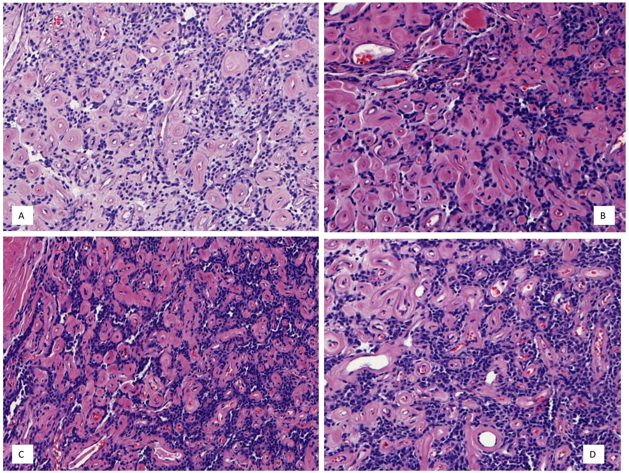
The wide application of increasingly advanced molecular studies in routine clinical practice has allowed a detailed, albeit still incomplete, genetic subclassification of undifferentiated round cell sarcomas. The WHO classification continues to include provisional molecular entities, whose clinicopathologic features are in the early stages of evolution. This review focuses on the clinicopathologic, molecular, and prognostic features of undifferentiated round cell sarcomas with EWSR1/FUS::NFATC2 or EWSR1::PATZ1 fusions. Classic histopathologic findings, uncommon variations, and diagnostic pitfalls are addressed, along with the utility of recently developed immunohistochemical and molecular markers.

Keywords: EWSR1::NFATC2 sarcoma; EWSR1::PATZ1 sarcoma; EWSR1::non-ETS sarcoma; Undifferentiated round cell sarcoma.


Figures

Figure 1.
Figure 1.
Magnetic resonance imaging (A) and macroscopic findings (B) of an EWSR1::NFATC2 sarcoma in fibula. (Courtesy Dra. Antonina Parafioriti, Milan).
Figure 2.
Figure 2.
A-C. EWSR1::NFATC2 sarcoma grows in lobules and sheets of round/oval to epithelioid cells with clear cytoplasm (hematoxylin and eosin (H&E) 200 and 400x). D. Cord-like pattern and chondromyxoid stromal tissue in an EWSR1::NFATC2 sarcoma (H&E 600x).
Figure 3.
Figure 3.
A-D. EWSR1::NFATC2 sarcoma arranged in anastomosing cords and clusters embedded within abundant myxohyaline stroma (H&E 100, 200 and 400x).
Figure 4.
Figure 4.
A-D. EWSR1::NFATC2 sarcoma arranged in clusters and pseudoacinar structures with hyaline and fibrous stroma (H&E 200 and 400x).
Figure 5.
Figure 5.
EWSR1::NFATC2 sarcoma with: A. poor stromal tissue and diffuse solid sheets of tumor cells, closely resembling a Ewing sarcoma (H&E 400x). B. poor stromal tissue and diffuse solid sheets of ovoid/spindle cells, closely resembling a BCOR-genetically altered sarcoma (H&E 400x) C. pleomorphic cells (H&E 400x). D. pseudoacinar structures and cord-like arrangement resembling a metastatic carcinoma (H&E 200x).
Figure 6.
Figure 6.
EWSR1::NFATc2 sarcoma with: A. CD99 expression with diffuse and membranous pattern. B. moderate NKX2.2 nuclear immunoreactivity. C. strong and diffuse PAX7 immunoreactivity D. Strong and diffuse ETV4 nuclear immunoexpression, which can raise the differential of CIC-rearranged sarcoma. E. strong and diffuse NKX3.1 immunoreactivity F. EWSR1 breakapart FISH analysis shows EWSR1 rearrangement and amplification in an EWSR1::NFATC2 sarcoma (100x) [Courtesy Dr Akihiko Yoshida, Tokyo].
Figure 7:
Figure 7:
A. Schematic illustration of the EWSR1::NFATC2 fusion. The two upper horizontal bars represent the genomic organization of the EWSR1 and NFATC2 genes. Blue and green solid boxes represent the exons (numbered), and the intron regions are lightly shaded. The arrows indicate the fusion points. The two horizontal bars in the lower segment of the figure show the genomic structure of the EWSR1::NFATC2 fusion gene. B. Schematic illustration of the EWSR1::PATZ1 fusion. The two upper horizontal bars represent the genomic organization of the EWSR1 and PATZ1 genes. Blue and green solid boxes represent the exons (numbered), and the intron regions are lightly shaded. The arrows indicate the fusion points and show that exons 8 and 9 of EWSR1 are fused to intra-exonic sequences in the first exon of PATZ1. The two horizontal bars in the lower segment of the figure show the genomic structure of the EWSR1::PATZ1 fusion genes.
Figure 8.
Figure 8.
A. Chest wall/periclavicular mass. B. Magnetic resonance imaging shows a large intramuscular mass in the left superior chest wall (trapezius muscle). C. EWSR1::PATZ1 sarcoma exhibiting well-circumscribed silhouette with variably thick fibrous capsule (H&E 200x and 400x).
Figure 9.
Figure 9.
A-D. EWSR1::PATZ1 sarcoma with multiple cystic structures and abundant fibrous stroma.
Figure 10.
Figure 10.
A-B. EWSR1::PATZ1 sarcoma with low-grade features composed of bland round/ovoid cells with low mitotic index, lack of necrosis and hyalinized/myxohyaline stromal tissue (H&E 200 and 400x)
Figure 11.
Figure 11.
A-B. EWSR1::PATZ1 sarcoma with low-grade features composed of round/ovoid cells with low mitotic index, hyalinized stromal tissue and extensive perimembranous collagen accentuation (H&E 200 and 400x).
Figure 12.
Figure 12.
A-D. EWSR1::PATZ1 sarcoma with undifferentiated round cell sarcoma morphology and minimal stroma (H&E 200, 400 and 600x).
Figure 13.
Figure 13.
A-D. EWSR1::PATZ1 sarcoma with undifferentiated round cell sarcoma morphology, exhibiting hyperchromatic nuclei and some bizarre forms, (H&E 400x)
Figure 14.
Figure 14.
EWSR1::PATZ1 sarcoma. A. patchy and nuclear S100 immunoreactivity. B. strong and diffuse GFAP positivity. C. focal and cytoplasmic desmin expression D. strong and diffuse MyoD1 immunoreactivity. E. dot-like CD99 expression. F. EWSR1 breakapart FISH demonstrating lack of a rearrangement with orange and green flanking probes remaining juxtaposed (less than a signal size apart).

References

    1. Antonescu C Round cell sarcomas beyond Ewing: emerging entities. Histopathology 2014;64:26–37. - PubMed
    1. Ohno T, Ouchida M, Lee L, et al. . The EWS gene, involved in Ewing family of tumors, malignant melanoma of soft parts and desmoplastic small round cell tumors, codes for an RNA binding protein with novel regulatory domains. Oncogene. 1994;9:3087–3097. - PubMed
    1. Tirode F, Surdez D, Ma X, et al. Genomic landscape of Ewing sarcoma defines an aggressive subtype with co-association of STAG2 and TP53 mutations. Cancer Discov. 2014;4:1342–53. - PMC - PubMed
    1. Crompton BD, Stewart C, Taylor-Weiner A, et al. The genomic landscape of pediatric Ewing sarcoma. Cancer Discov. 2014;4:1326–41. - PubMed
    1. Mantilla JG, Ricciotti RW, Chen E, et al. Detecting disease-defining gene fusions in unclassified round cell sarcomas using anchored multiplex PCR/targeted RNA next-generation sequencing-Molecular and clinicopathological characterization of 16 cases. Genes Chromosomes Cancer. 2019;58:713–722. - PubMed