. 2024 Jan 26;15(1):793.
doi: 10.1038/s41467-024-44967-z.

The transcription factor AP2XI-2 is a key negative regulator of Toxoplasma gondii merogony

Affiliations

The transcription factor AP2XI-2 is a key negative regulator of Toxoplasma gondii merogony

Jin-Lei Wang et al. Nat Commun. .

Abstract

Sexual development in Toxoplasma gondii is a multistep process that culminates in the production of oocysts, constituting approximately 50% of human infections. However, the molecular mechanisms governing sexual commitment in this parasite remain poorly understood. Here, we demonstrate that the transcription factors AP2XI-2 and AP2XII-1 act as negative regulators, suppressing merozoite-primed pre-sexual commitment during asexual development. Depletion of AP2XI-2 in type II Pru strain induces merogony and production of mature merozoites in an alkaline medium but not in a neutral medium. In contrast, AP2XII-1-depleted Pru strain undergoes several rounds of merogony and produces merozoites in a neutral medium, with more pronounced effects observed under alkaline conditions. Additionally, we identified two additional AP2XI-2-interacting proteins involved in repressing merozoite programming. These findings underscore the intricate regulation of pre-sexual commitment by a network of factors and suggest that AP2XI-2 or AP2XII-1-depleted Pru parasites can serve as a model for studying merogony in vitro.


Conflict of interest statement

The authors declare no competing interests.

Figures
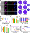
Fig. 1
Fig. 1. AP2XI-2 is critical for the growth of Toxoplasma gondii in vitro and in vivo.
a AP2XI-2-mAID-6HA is localized to the nucleus in the tachyzoites (32 h post-infection) and bradyzoites (3 days post-infection) and is efficiently depleted when the parasites are treated with IAA (3-indoleacetic acid). Magenta, anti-IMC1; green, FITC-Dolichos biflorus lectin (FITC-DBL); red, anti-HA. Scale bar, 10 µm. b Representative images of the plaques formed by the indicated parasite strains grown in HFF monolayers for 9 days. c Relative size of the plaques detected in b. Data represents the mean ± SD from three independent experiments. Statistical significance was tested by two-tailed, unpaired t test, ****p < 0.0001. d Quantification of invasion of the indicated strains grown in HFF monolayers in the presence or absence of IAA. Data represents the mean ± SD from three independent experiments, analyzed by two-tailed, unpaired t test, *p = 0.0234. e Quantification of the replication of the indicated strains grown in HFFs for 30 h in the presence or absence of IAA (added 2 h post-invasion). Data represents the mean ± SD from three independent experiments, analyzed by two-way ANOVA with Tukey multiple comparison test, ****p < 0.0001. f Quantification of the egress of the indicated strains grown in HFFs for 48-60 h in the presence or absence of IAA (added 2 h post-invasion). Data represents the mean ± SD from three independent experiments, analyzed by two-tailed, unpaired t test, *p = 0.0365. g Survival of C57BL/6 mice infected by 2 × 104 or 2 × 105 tachyzoites of the indicated strains. Survival curves of mice were compared using Gehan-Breslow-Wilcoxon test, p < 0.0001. (n = 15 mice/strain for the 2 × 104 dose; n = 10 mice/strain for the 2 × 105 dose). h The average body weight ± SD of the mice infected by 2 × 105 of the parental Pru or PruΔap2XI-2 tachyzoites. (n = 10 mice / strain). Source data are provided as a Source data file.
Fig. 2
Fig. 2. Loss of AP2XI-2 blocks bradyzoite differentiation and results in severe morphological abnormalities under alkaline conditions.
a Representative images of the indicated parasite strains grown in an alkaline culture medium without CO2 for 3 days for induction of bradyzoites. Red, anti-IMC1; green, FITC-DBL; magenta, anti-BAG1. Scale bar, 10 µm. b Quantification of bradyzoite differentiation in the indicated strains 3 days after exposure to alkaline stress. Data are presented as the mean ± SD from at least three independent biological replicates. The percentage of DBL-positive vacuoles was calculated based on at least 150 vacuoles per replicate. The percentage of vacuoles scoring “DBL-high” was used for comparison and analyzed by two-tailed, unpaired t test, ***p = 0.0002 (AP2XI-2-mAID -IAA vs +IAA), ***p = 0.0001 (Pru vs PruΔap2XI-2). c Heat map showing the decreased expression of selected bradyzoite highly expressed genes after depletion of AP2XI-2 under alkaline conditions. The color scale indicates log10-transformed FPKM values. Source data are provided as a Source data file.
Fig. 3
Fig. 3. Depletion of AP2XI-2 induces tachyzoites-to-merozoites switch under alkaline condition.
a Volcano plot showing the differentially expressed genes in the AP2XI-2-expressing and AP2XI-2-depleted strains grown under alkaline culture conditions for 3 days. The merozoite, bradyzoite, sporozoite and tachyzoite highly expressed genes were analyzed from Supplementary data 1. Data from four biological replicates were plotted and a fold change of ≥2.0 or ≤−2.0 with a p-value < 0.05 was considered statistically significant. b Volcano plot showing the differentially expressed genes in the AP2XI-2-expressing and AP2XI-2-depleted strains grown under neutral culture conditions. The merozoite, bradyzoite, sporozoite and tachyzoite highly expressed genes were analyzed from Supplementary data 1. Data from four biological replicates were plotted and a fold change of ≥2.0 or ≤−2.0 with a p-value < 0.05 was considered statistically significant. c Representative images of AP2XI-2 knockdown (KD) parasite strains grown in a neutral or an alkaline culture medium without CO2 for 3 days for induction of merogony. IMC1 (magenta) specifically stains the (developing) merozoites, whereas IMC7-2Ty (red) is specific to the polyploid AP2XI-2-depleted schizonts. M merozoite, S schizont. Scale bar, 10 µm.
Fig. 4
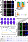
Fig. 4. AP2XII-1, an interacting partner of the AP2XI-2 complex, also silences merozoite-primed pre-sexual commitment.
a AP2XII-1-mAID-6HA localizes to the nucleus in tachyzoites (32 h post-infection) and bradyzoites (3 days post-infection) and is efficiently depleted when the parasites are treated with IAA. Magenta, anti-IMC1; green, FITC-DBL; red, anti-HA. Scale bar, 10 µm. b Quantification of bradyzoite differentiation in the indicated strains following 3 days of exposure to an alkaline culture medium. Data are presented as the mean ± SD from at least three independent biological replicates and the percentage of DBL-positive vacuoles was calculated based on a minimum of 150 vacuoles per replicate. The percentage of vacuoles scoring “DBL-high” was used for comparison and analyzed by two-tailed, unpaired t test, *p = 0.0379 (AP2XII-1-mAID, -IAA vs +IAA), **p = 0.0020 (209500-mAID, -IAA vs +IAA), ***p = 0.0003 (214140-mAID, -IAA vs +IAA), *p = 0.0449 (275680-mAID, -IAA vs +IAA), **p = 0.0022 (TIR1 -IAA vs 275680-mAID -IAA), ****p < 0.0001 (TIR1 -IAA vs AP2XII-1-mAID -IAA). c Representative images of the plaques formed by the indicated parasites grown in HFF monolayers for 8 days. d Relative size of the plaques detected in c. Data represents the mean ± SD from three independent experiments. Statistical significance was tested by two-tailed, unpaired t test, **p = 0.0019 (TIR1 -IAA vs AP2XII-1-mAID -IAA), **p = 0.0074 (209500-mAID -IAA vs +IAA), **p = 0.0014 (214140-mAID -IAA vs +IAA), **p = 0.0067 (TIR1 -IAA vs 275680-mAID -IAA), ****p < 0.0001. e The subcellular location of the indicated proteins in the mature merozoites. The indicated parasites were allowed to infect HFFs for 4 h followed by incubation in an alkaline medium without CO2 for 3 days with IAA. Green, anti-IMC1; red, anti-Ty; Scale bar, 10 µm. f Genome-wide analysis reveals that AP2XI-2, AP2XII-1 and MORC associate with the transcriptional start sites (TSS) of the genes. The indicated parasites treated with or without IAA for 48 h were used to assess the genomic loci of AP2XI-2, AP2XII-1 or MORC. g Integrated genome browser view of AP2XI-2, AP2XII-1 and MORC enrichment across T. gondii chromosome Ia. The data of MORC-HA Rep1 and MORC-HA Rep2 were obtained from the GEO under accession number GSE136060. Source data are provided as a Source data file.
Fig. 5
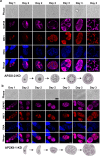
Fig. 5. Characterization of merozoite formation after depletion of AP2XI-2 or AP2XII-1 under alkaline conditions.
Representative images of AP2XI-2 (a) or AP2XII-1 (b) knock-down (KD) parasites grown in an alkaline medium without CO2 over 3 days for induction of merozoites. Magenta, anti-GAP45; red, anti-IMC1. Scale bar, 10 µm.
Fig. 6
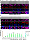
Fig. 6. Characterization of the roles of AP2XI-2 interacting proteins in the AP2XI-2-depleted parasites.
a, b Representative images of AP2XI-2 and AP2XII-1 co-depleted parasites grown in neutral (a) or alkaline medium (b) over 3 days for induction of merozoites. Magenta, anti-GAP45; red, anti-IMC1. Scale bar, 10 µm. c Quantitative analysis of the budding patterns in the indicated strains treated with IAA for 3 days. Data are presented as the mean ± SD from three independent biological replicates. The percentage of zoites per maternal parasite was calculated based on at least 50 parasites per replicate. Statistical significance was tested by two-tailed, unpaired t test. *Compared with the percentage of >16 zoites in the AP2XI-2-KD cultured under an alkaline medium, ****p < 0.0001; #compared with the percentage of 8–16 zoites in the AP2XII-1-KD cultured under a neutral medium, #p = 0.016 for AP2XII-1-KD cultured under an alkaline medium; ##p = 0.0024 for AP2XI-2-KD-AP2XII-1-KD cultured under a neutral medium; ##p = 0.0013 for AP2XI-2-KD-AP2XII-1-KD cultured under an alkaline medium.
Fig. 7
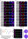
Fig. 7. Several secondary transcriptional regulators are activated to regulate developmental stage transition after depletion of AP2XI-2.
a Confocal images showing the expression of secondary transcriptional regulators during merogony after depletion of AP2XI-2 under alkaline conditions for 3 days. The secondary transcriptional AP2 factors shown in blue and green colors on the left side of the figure denote those mainly expressed in mature merozoites and schizonts, respectively. Factors shown in black color denote those with slight increase upon depletion of AP2XI-2. Magenta, anti-IMC1; red, anti-Ty. Scale bar, 10 µm. b Representative images of the indicated parasite strains grown in an alkaline medium for 3 days for induction of merozoites. Magenta, anti-GAP45; red, anti-IMC1. Scale bar, 10 µm. c Quantification of mature merozoites in the indicated strains following 3 days of exposure to alkaline stress. Data represents the mean ± SD from three independent experiments, analyzed by two-tailed, unpaired t test, NS, no significant. d AP2X-3-mAID-6HA was undetected in tachyzoites (32 h post-infection) and bradyzoites (3 days post-infection). Magenta, anti-IMC1; green, FITC-DBL; red, anti-HA. Scale bar, 10 µm. e Representative images of the plaques formed by AP2X-3-mAID-6HA parasites grown in HFF monolayers for 8 days with or without IAA.

References

    1. Dubey JP, Lindsay DS, Speer CA. Structures of Toxoplasma gondii tachyzoites, bradyzoites, and sporozoites and biology and development of tissue cysts. Clin. Microbiol. Rev. 1998;11:267–299. doi: 10.1128/CMR.11.2.267. - DOI - PMC - PubMed
    1. Montoya JG, Liesenfeld O. Toxoplasmosis. Lancet. 2004;363:1965–1976. doi: 10.1016/S0140-6736(04)16412-X. - DOI - PubMed
    1. Robert-Gangneux F, Dardé ML. Epidemiology of and diagnostic strategies for toxoplasmosis. Clin. Microbiol. Rev. 2012;25:264–296. doi: 10.1128/CMR.05013-11. - DOI - PMC - PubMed
    1. Elsheikha HM, Marra CM, Zhu XQ. Epidemiology, pathophysiology, diagnosis, and management of cerebral toxoplasmosis. Clin. Microbiol. Rev. 2021;34:e00115–19. - PMC - PubMed
    1. Kim K. The epigenome, cell cycle, and development in Toxoplasma. Annu. Rev. Microbiol. 2018;72:479–499. doi: 10.1146/annurev-micro-090817-062741. - DOI - PubMed