. 2024 Jan 26;14(1):2240.
doi: 10.1038/s41598-024-52689-x.

Bezafibrate attenuates immobilization-induced muscle atrophy in mice

Affiliations

Bezafibrate attenuates immobilization-induced muscle atrophy in mice

Satoshi Nakamura et al. Sci Rep. .

Abstract

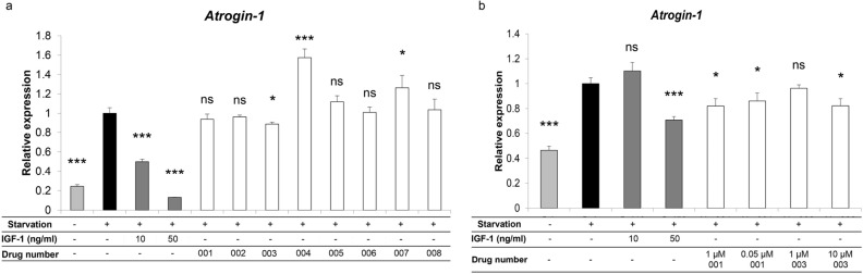
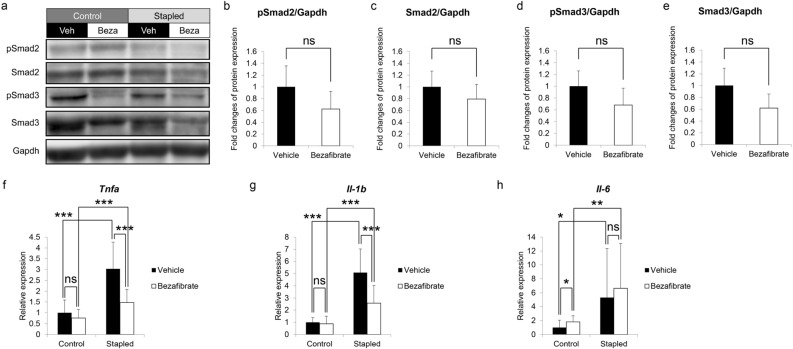
Muscle atrophy due to fragility fractures or frailty worsens not only activity of daily living and healthy life expectancy, but decreases life expectancy. Although several therapeutic agents for muscle atrophy have been investigated, none is yet in clinical use. Here we report that bezafibrate, a drug used to treat hyperlipidemia, can reduce immobilization-induced muscle atrophy in mice. Specifically, we used a drug repositioning approach to screen 144 drugs already utilized clinically for their ability to inhibit serum starvation-induced elevation of Atrogin-1, a factor related to muscle atrophy, in myotubes in vitro. Two candidates were selected, and here we demonstrate that one of them, bezafibrate, significantly reduced muscle atrophy in an in vivo model of muscle atrophy induced by leg immobilization. In gastrocnemius muscle, immobilization reduced muscle weight by an average of ~ 17.2%, and bezafibrate treatment prevented ~ 40.5% of that atrophy. In vitro, bezafibrate significantly inhibited expression of the inflammatory cytokine Tnfa in lipopolysaccharide-stimulated RAW264.7 cells, a murine macrophage line. Finally, we show that expression of Tnfa and IL-1b is induced in gastrocnemius muscle in the leg immobilization model, an activity significantly antagonized by bezafibrate administration in vivo. We conclude that bezafibrate could serve as a therapeutic agent for immobilization-induced muscle atrophy.


Conflict of interest statement

The authors declare no competing interests.

Figures

Figure 1
Figure 1
Effects of existing drugs on serum starvation-induced Atrogin-1 expression in C2C12 myoblasts. (a) C2C12 cells were cultured for 72 h in DMEM with 2% horse serum to induce myotube formation. Subsequently, myotubes were cultured with or without (serum starvation) 2% horse serum for 24 h and stimulated with indicated drugs (each 1 µM) or 10 or 50 ng/ml rhIGF-1 for the last 6 h. Atrogin-1 expression was analyzed by realtime PCR. Data represent mean Atrogin-1 expression relative to Gapdh ± SD. Serum starvation without drugs or IGF-1 served as controls (each n = 3, *P < 0.05, ***P < 0.001 by Student’s t-test). (b) C2C12 cells were cultured 24 h in DMEM with or without (serum starvation) 10% FBS in the presence or absence of indicated concentrations of drugs No.001 or 003 for the last 6 h. Atrogin-1 expression was then analyzed by realtime PCR. Data represent mean Atrogin-1 expression relative to Gapdh ± SD. Serum starvation without drugs served as controls (each n = 3, *P < 0.05, ***P < 0.001 by Student’s t-test).
Figure 2
Figure 2
Bezafibrate reduces immobilization- but not denervation-induced muscle atrophy in mice. Nine-week-old C57BL/6 mice were treated with No.001, No.003 (bezafibrate) or vehicle once daily for eight days, and staple fixation or sciatic nerve denervation surgery (DEN) was performed on left hind limbs on day two. On day nine, mice were sacrificed and gastrocnemius and quadriceps muscles were harvested. Right hind limbs served as controls (sham-operated) (n = 5, each group). Wet weights of gastrocnemius (a,c) and quadriceps (b,d) muscle adjusted to body weight in the stapled plus drug conditions (No.001, (a,b); No.003, (c,d)) versus the control side were analyzed. Wet weights of gastrocnemius (e) and quadriceps (f) muscle adjusted to body weight in the denervated plus No.003 condition versus the sham side are shown. Representative data of two independent experiments are shown. (*P < 0.05; ***P < 0.001 by Student’s t-test). (gj) Relative mean cross-sectional areas (CSA) of gastrocnemius (g) and quadriceps (i) muscles from mice treated with bezafibrate, with or without staple fixation. (**P < 0.01; ***P < 0.001 by Student’s t-test). Boxplot of CSA of gastrocnemius (h) and quadriceps (j) muscles from same mice. CSA was measured at three randomly selected regions. The number of myofibers evaluated per mouse in the vehicle group was 178–294 or 132–401 in gastrocnemius or quadriceps, respectively. In the bezafibrate group it was 164–266 or 134–230 in gastrocnemius or quadriceps. (gj). (k,l) Hematoxylin and eosin staining of gastrocnemius (k) and quadriceps (l) muscles. Scale bar, 100 µm.
Figure 3
Figure 3
Bezafibrate treatment decreases Tnfa and Il-1b expression but does not inhibit Smad2/3 protein accumulation or phosphorylation in stapled gastrocnemius muscles. (ae) Nine-week-old C57BL/6 mice were treated with bezafibrate or vehicle once daily for eight days. Staple fixation was performed on left hind limbs on day two of drug treatment. On day nine, mice were sacrificed and gastrocnemius muscles were harvested. Accumulation of Smad2, phosphorylated Smad2 (pSmad2), Smad3 and phosphorylated Smad3 (pSmad3) proteins in same muscles, as detected by western blotting (a). Representative images are shown. Accumulation of pSmad2 (b), Smad2 (c), pSmad3 (d), and Smad3 (e) protein on the fixed side of gastrocnemius muscle based on ratios of indicated proteins to Gapdh protein, based on ImageJ. Data represent mean ± SD, and vehicle groups served as controls (each group n = 3. ns; not significant by Student’s t-test). Relative expression of Tnfa (f), Il-1b (g) and Il-6 (h) in the same muscles, based on realtime PCR (fh). Data represent mean gene expression relative to Gapdh ± SD (each n = 5, *P < 0.05; **P < 0.01; ***P < 0.001 by Student’s t-test).
Figure 4
Figure 4
LPS-induced Tnfa expression in macrophages decreases after bezafibrate treatment. (a) RAW 264.7 cells were cultured 24 h with or without 10 ng/ml LPS in the presence or absence of indicated concentrations of bezafibrate. Relative Tnfa expression was then analyzed by realtime PCR. Data represent mean Tnfa expression relative to Gapdh ± SD (each n = 3, *P < 0.05; **P < 0.01; ***P < 0.001 by Student’s t-test). (b) Nine-week-old C57BL/6 mice were treated with bezafibrate or vehicle once daily for eight days. Staple fixation was performed on left hind limbs on day two of drug treatment. On day nine, mice were sacrificed and gastrocnemius muscles were harvested. Specimens were stained with Alexa Fluor 488-conjugated rat anti-mouse F4/80 and rabbit anti-mouse TNFα, followed by Alexa546-conjugated goat anti-Rabbit IgG. Nuclei were stained with DAPI. Sections were observed under a fluorescence microscope and all magnifications are 400x. Scale bar, 100 µm. In merged samples, images shown in the rightmost column are higher magnifications of squared areas in the adjacent image. In the latter, scale bars indicate 50 µm. (c) Shown is the ratio of the number of F4/80/TNFα double-positive cells and the number of DAPI-positive nuclei measured at five randomly selected locations. Data represent mean ratios relative to numbers observed in the vehicle (control) group ± SD (n = 5, *P < 0.05; **P < 0.01; ***P < 0.001 by one-way ANOVA and Tukey post hoc test).

Similar articles

References

    1. Han HQ, Zhou X, Mitch WE, Goldberg AL. Myostatin/activin pathway antagonism: molecular basis and therapeutic potential. Int. J. Biochem. Cell Biol. 2013;45:2333–2347. doi: 10.1016/j.biocel.2013.05.019. - DOI - PubMed
    1. Lecker SH, Goldberg AL, Mitch WE. Protein degradation by the ubiquitin-proteasome pathway in normal and disease states. J. Am. Soc. Nephrol. 2006;17:1807–1819. doi: 10.1681/asn.2006010083. - DOI - PubMed
    1. Costelli P, et al. Ca(2+)-dependent proteolysis in muscle wasting. Int. J. Biochem. Cell Biol. 2005;37:2134–2146. doi: 10.1016/j.biocel.2005.03.010. - DOI - PubMed
    1. Sartori R, Romanello V, Sandri M. Mechanisms of muscle atrophy and hypertrophy: Implications in health and disease. Nat. Commun. 2021;12:330. doi: 10.1038/s41467-020-20123-1. - DOI - PMC - PubMed
    1. Bodine SC, et al. Identification of ubiquitin ligases required for skeletal muscle atrophy. Science (New York) 2001;294:1704–1708. doi: 10.1126/science.1065874. - DOI - PubMed