Case Reports
. 2024 Jun;35(2):122-133.
doi: 10.1007/s12022-024-09798-0. Epub 2024 Jan 27.

Spatial Transcriptomics in a Case of Follicular Thyroid Carcinoma Reveals Clone-Specific Dysregulation of Genes Regulating Extracellular Matrix in the Invading Front

Affiliations
Case Reports

Spatial Transcriptomics in a Case of Follicular Thyroid Carcinoma Reveals Clone-Specific Dysregulation of Genes Regulating Extracellular Matrix in the Invading Front

Vincenzo Condello et al. Endocr Pathol. 2024 Jun.

Abstract

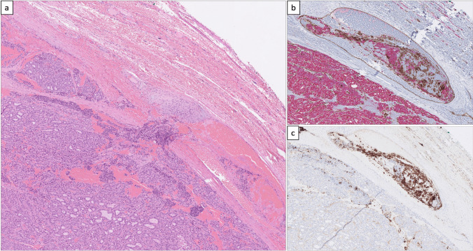
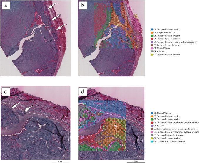
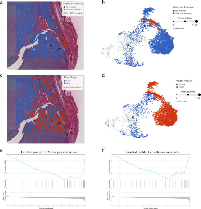
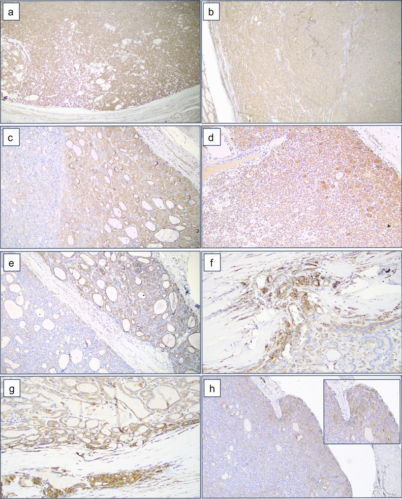
Follicular thyroid carcinoma (FTC) is recognized by its ability to invade the tumor capsule and blood vessels, although the exact molecular signals orchestrating this phenotype remain elusive. In this study, the spatial transcriptional landscape of an FTC is detailed with comparisons between the invasive front and histologically indolent central core tumor areas. The Visium spatial gene expression platform allowed us to interrogate and visualize the whole transcriptome in 2D across formalin-fixated paraffin-embedded (FFPE) tissue sections. Four different 6 × 6 mm areas of an FTC were scrutinized, including regions with capsular and vascular invasion, capsule-near area without invasion, and a central core area of the tumor. Following successful capturing and sequencing, several expressional clusters were identified with regional variation. Most notably, invasive tumor cell clusters were significantly over-expressing genes associated with pathways interacting with the extracellular matrix (ECM) remodeling and epithelial-to-mesenchymal transition (EMT). Subsets of these genes (POSTN and DPYSL3) were additionally validated using immunohistochemistry in an independent cohort of follicular thyroid tumors showing a clear gradient pattern from the core to the periphery of the tumor. Moreover, the reconstruction of the evolutionary tree identified the invasive clones as late events in follicular thyroid tumorigenesis. To our knowledge, this is one of the first 2D global transcriptional mappings of FTC using this platform to date. Invasive FTC clones develop in a stepwise fashion and display significant dysregulation of genes associated with the ECM and EMT - thus highlighting important molecular crosstalk for further investigations.

Keywords: FTC; Invasion; Phylogenetics, Extracellular matrix; Spatial transcriptomics.


Conflict of interest statement

The authors declare no competing interests.

Figures

Fig. 1
Fig. 1
Histological attributes of the index case. a Hematoxylin-eosin-stained section depicting extensive vascular invasion. b Double stain (CD31 in brown, pan-cytokeratin in red) visualizing intravascular tumor deposits. c CD61 immunohistochemistry confirms an in vivo phenomenon with thrombus
Fig. 2
Fig. 2
Outline of the spatial transcriptomics analysis. Tiled versions of the two tissue images showing invasion that were used for spatial transcriptomics analysis are shown in ad. a Hematoxylin-eosin-stained section of the vascular invasion area; arrows indicate the invading foci. b Unsupervised clustering analysis showing nine different clusters in slide 1 with vascular invasion. Each color represents a distinct cluster. c Hematoxylin-eosin-stained section of the capsular invasion area; arrows indicate the invading foci. d Unsupervised clustering analysis showing 10 different clusters in slide 2 with capsular invasion. Each color represents a distinct cluster (C1–10). Each spot within the 6 × 6-mm square represents an area with gene expression information, allowing for gene expression–based clustering information
Fig. 3
Fig. 3
Trajectory analysis and gene set enrichment analysis in the angioinvasive tissue sample. a Hematoxylin-eosin-stained section of angioinvasive tissue with filtered tumor cell spots for analysis. b Trajectory analysis with tumor cell spots, showing clustering of angioinvasive tumor cell spots occurring with high pseudotime. c Hematoxylin-eosin-stained section of angioinvasive tissue representing the time stage of “early” and “late” tumor cells. d Trajectory analysis dichotomized into “early” and “late” tumor cells based on median pseudotime. e Gene Set Enrichment Analysis (GSEA) plots demonstrating downregulation of ECM-receptor interaction (path:hsa04512) in early versus late (NES −2.03, p-value 0.00, FDR 0.00). f Gene set enrichment analysis (GSEA) plots showing the downregulation of cell adhesion molecules (path:hsa04514) in early versus late (NES −0.51, p-value 0.00, FDR 0.05)
Fig. 4
Fig. 4
Extracellular matrix (ECM) marker immunohistochemistry. Immunohistochemical staining of DPYSL3 and POSTN in follicular thyroid carcinoma (FTC) compared to follicular thyroid adenoma (FTA). a, b Staining showing the homogeneous expression of DPYSL3 and POSTN respectively in FTA. c, d Pronounced expression of DPYSL3 and POSTN, respectively, characterized by increased intensity extending from the central region towards the periphery of the FTC. e, f Strong expression of DPYSL3 and POSTN respectively in the area with capsular invasion. Statistical significance for this feature was observed only for DPYSL3. g, h Pronounced expression of DPYSL3 and POSTN in the area of vascular invasion. Statistical significance for this feature was observed only for DPYSL3

Similar articles

Cited by

References

    1. D’Avanzo A, Treseler P, Ituarte PHG, Wong M, Streja L, Greenspan FS, et al. Follicular thyroid carcinoma: Histology and prognosis. Cancer. 2004;100:1123–9. doi: 10.1002/cncr.20081. - DOI - PubMed
    1. Aschebrook-Kilfoy B, Grogan RH, Ward MH, Kaplan E, Devesa SS. Follicular Thyroid Cancer Incidence Patterns in the United States, 1980–2009. Thyroid. 2013;23:1015–21. doi: 10.1089/thy.2012.0356. - DOI - PMC - PubMed
    1. Aschebrook-Kilfoy B, Ward MH, Sabra MM, Devesa SS. Thyroid Cancer Incidence Patterns in the United States by Histologic Type, 1992–2006. Thyroid. 2011;21:125–34. doi: 10.1089/thy.2010.0021. - DOI - PMC - PubMed
    1. WHO Classification of Tumours Editorial Board: Endocrine and Neuroendocrine tumours. vol. 8. 5th ed. Lyon, France: International Agency for Research on Cancer; 2022.
    1. Nikiforov YE, Seethala RR, Tallini G, Baloch ZW, Basolo F, Thompson LDR, et al. Nomenclature Revision for Encapsulated Follicular Variant of Papillary Thyroid Carcinoma: A Paradigm Shift to Reduce Overtreatment of Indolent Tumors. JAMA Oncol. 2016;2:1023. doi: 10.1001/jamaoncol.2016.0386. - DOI - PMC - PubMed

Publication types

MeSH terms