. 2024 Mar 15;5(1):102840.
doi: 10.1016/j.xpro.2024.102840. Epub 2024 Jan 26.

Analyzing engram reactivation and long-range connectivity

Affiliations

Analyzing engram reactivation and long-range connectivity

Ron Refaeli et al. STAR Protoc. .

Abstract

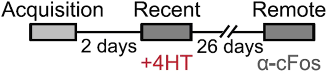
Here, we present a protocol for marking engram cells to efficiently measure reactivation levels and their projection pathways. We describe steps for genetic manipulation utilizing transgenic mice and viral infections, labeling engram cells, and a modified version of CLARITY, a tissue-clearing technique. This protocol can be adapted to various research inquiries that involve assessing the overlap of cell populations and uncovering novel long-range connectivity pathways. For complete details on the use and execution of this protocol, please refer to Refaeli et al. (2023).1.

Keywords: Behavior; Microscopy; Model Organisms; Neuroscience.


Conflict of interest statement

Declaration of interests The authors declare no competing interests.

Figures

None
Graphical abstract
Figure 1
Figure 1
Scheme of the behavioral paradigm used in this protocol Mice were trained in Contextual fear conditioning and recall this aversive memory after two days and 28 days. Both recall events were followed by engram group tagging, the first in-vivo, and the second by IHC. Any other behavioral paradigm with two time points can be performed similarly.
Figure 2
Figure 2
Image analysis using Imaris (A) The ‘Surface’ option on Imaris is used to define boundaries. The green arrow indicates the option to download the desired excel data. (B) The ‘Spots’ option used to define somata position. (C) TdTomato+ cells in the pyramidal layer of the CA1. (D) Marking the somata of all red cells using the Spots option.
Figure 3
Figure 3
Polymerizing the brain This step involves immersing the fixed tissue in a hydrogel solution and substituting all oxygen with nitrogen. For multiple samples, nitrogen can be replaced with oxygen simultaneously in several tubes (optional). The brain and hydrogel solution around it are then heated to enable polymerization. Over time, the tissue and the surrounding solution combine to form a monomer.
Figure 4
Figure 4
Chamber preparation (A) Preparing hot glue walls ensuring ample space for the tissue and leaving a small gap (upper left corner). (B) Constructing the walls to match the height of the tissue. After sealing the chamber with a coverslip (C), it can be easily filled with CSRC (D). (E) Closing the gap through which the CSRC was filled. (F) Once the chamber is sealed, an additional layer of hot glue is added above the coverslip to contain the immersion liquid. The tissue exhibits sufficient transparency for imaging under the microscope.
Figure 5
Figure 5
Tracing single neurons and single axons inside an entire clear brain (A) Engram cells at the dorsal CA1 region of the hippocampus of a cleared brain express TdTomato (red). Projections pathways can be easily targeted, heading toward the ACC (arrow). Scale bar = 700 μm. (B) An entire brain was imaged, at a resolution sufficient to separate and count single axons when magnified. Scale bar = 100 μm. (C) Projections can be explored throughout the entire brain. As shown here, this bundle of axons reaches the mammillary bodies (dashed line) via the Fornix (arrow). Scale bar = 300 μm. (D) Using CLARITY allows answering exploratory questions regarding the brain. i.e., not just ‘does the CA1 interact with the ACC?’ but ‘toward which areas does the CA1 sends projections?’ Scale bar = 700 μm.
Figure 6
Figure 6
Examples of brain images using different methods (A) Thin slice of the CA1 section of Ai14 reporter mice hippocampus infected with the AAV5-c-Fos::CreER vector. Each red cell (TdTomato+) expressed c-Fos when the 4-OHT was introduced. When the drug administration is timed to the behavioral paradigm, those cells can be referred to as the engram cells of that behavioral task. (B) The CA1 section of the hippocampus in Ai14 mouse infected with AAV5-c-Fos::CreER, all red cells (TdTomato+) represent the engram cells of the first time point. All green cells (GFP+) are CA1→ACC projecting cells, due to injection of the retrograde vector to the ACC (AAVretro-CaMKII::sGFP), and all white cells represent the engram population of the second time point, after staining against the c-Fos protein (AF 647), adjacent to the memory task. (C) TdTomato+ cells in thick sample. Using the CLARITY protocol, a large portion of the dorsal CA1 can be easily imaged under a confocal microscope, to allow 3D information of the chosen engram population. (D) The use of our modified CLARITY protocol allows imaging of an entire brain, at a resolution of single cells. From above, the hippocampus is visible (green channel) along with every cell projecting to the ACC, due to infection with AAVretro-CaMKII::GFP.
Figure 7
Figure 7
After the chamber is created, aggregates may form in the brain (A) Transparent brain in a chamber. (B) Aggregates appear in the brain, whitening it and harming its structure.

References

    1. Refaeli R., Kreisel T., Groysman M., Adamsky A., Goshen I. Engram stability and maturation during systems consolidation. Curr. Biol. 2023;25:3942–3950. - PMC - PubMed
    1. Madisen L., Zwingman T.A., Sunkin S.M., Oh S.W., Zariwala H.A., Gu H., Ng L.L., Palmiter R.D., Hawrylycz M.J., Jones A.R., et al. A robust and high-throughput Cre reporting and characterization system for the whole mouse brain. Nat. Neurosci. 2010;13:133–140. - PMC - PubMed
    1. Refaeli R., Goshen I. Investigation of Spatial Interaction Between Astrocytes and Neurons in Cleared Brains. J. Vis. Exp. 2022 - PubMed
    1. Ye L., Allen W.E., Thompson K.R., Tian Q., Hsueh B., Ramakrishnan C., Wang A.C., Jennings J.H., Adhikari A., Halpern C.H., et al. Wiring and Molecular Features of Prefrontal Ensembles Representing Distinct Experiences. Cell. 2016;165:1776–1788. - PMC - PubMed
    1. Du H., Hou P., Zhang W., Li Q. Advances in CLARITY-based tissue clearing and imaging. Exp. Ther. Med. 2018;16:1567–1576. - PMC - PubMed

Publication types

LinkOut - more resources