. 2024 Apr 15;75(8):2313-2329.
doi: 10.1093/jxb/erae031.

Functional characterization of calmodulin-like proteins, CML13 and CML14, as novel light chains of Arabidopsis class VIII myosins

Affiliations

Functional characterization of calmodulin-like proteins, CML13 and CML14, as novel light chains of Arabidopsis class VIII myosins

Kyle Symonds et al. J Exp Bot. .

Abstract

Myosins are important motor proteins that associate with the actin cytoskeleton. Structurally, myosins function as heteromeric complexes where smaller light chains, such as calmodulin (CaM), bind to isoleucine-glutamine (IQ) domains in the neck region to facilitate mechano-enzymatic activity. We recently identified Arabidopsis CaM-like (CML) proteins CML13 and CML14 as interactors of proteins containing multiple IQ domains, including a myosin VIII. Here, we demonstrate that CaM, CML13, and CML14 bind the neck region of all four Arabidopsis myosin VIII isoforms. Among CMLs tested for binding to myosins VIIIs, CaM, CML13, and CML14 gave the strongest signals using in planta split-luciferase protein interaction assays. In vitro, recombinant CaM, CML13, and CML14 showed specific, high-affinity, calcium-independent binding to the IQ domains of myosin VIIIs. CaM, CML13, and CML14 co-localized to plasma membrane-bound puncta when co-expressed with red fluorescent protein-myosin fusion proteins containing IQ and tail domains of myosin VIIIs. In vitro actin motility assays using recombinant myosin VIIIs demonstrated that CaM, CML13, and CML14 function as light chains. Suppression of CML13 or CML14 expression using RNA silencing resulted in a shortened-hypocotyl phenotype, similar to that observed in a quadruple myosin mutant, myosin viii4KO. Collectively, our data indicate that Arabidopsis CML13 and CML14 are novel myosin VIII light chains.

Keywords: Calmodulin-like proteins; cytoskeleton; motor protein; myosin light chains; plant myosins.


Conflict of interest statement

The authors have no conflicts to declare.

Figures

Fig. 1.
Fig. 1.
Protein sequence alignments of (A) the neck regions (residues ~800–1000) for Arabidopsis class VIII myosins, and (B) Arabidopsis CaM (AtCaM7, AT3G43810), CML13, and CML14. Amino acid residues were shaded based on their percentage identity, dark gray if identical, and progressively lighter gray through to white as unconserved. In (A), predicted IQ motif consensus sequences are overlined. The EF-hands of CaM in (B) are overlined, and Ca2+-coordinating residues in CaM are marked with a dot beneath the alignment. Residues that differ between CML13 and CML14 are indicated with an asterisk. ClustalΩ was used for alignment (Sievers and Higgins, 2014; Gouy et al., 2021), and images were generated using Jalview Version 2.11.2.6. (C) Phylogenetic tree of Arabidopsis class VIII and XI myosins. The tree was constructed using Seaview version 5.0 (Sievers and Higgins, 2014; Gouy et al., 2021) from a complete alignment of proteins using PhyML with bootstrapping analysis (1000 replicates).
Fig. 2.
Fig. 2.
Split-luciferase protein interaction of Arabidopsis CMLs with the neck region of class VIII myosins in planta. N. benthamiana leaves were infiltrated with Agrobacterium harboring respective NLuc–prey (myosin VIII neck regions) and CLuc–bait (CaM, CMLs) vectors and tested for luciferase activity 4 d after inoculation, as described in the Materials and methods. (A) Schematic representation of myosin VIIIs showing the relative positions of myosin domains. In planta analysis of various CMLs with the neck regions of myosin (B) ATM1 (residues 848–943), (C) ATM2 (residues 878–967), (D) VIII-A (residues 828–930), and (E) VIII-B (residues 834–913), respectively. Split-luciferase data are expressed as a fold difference relative to the RLU signal (log10 scale) observed using the negative control bait CML42 which was set to an RLU of 1.0. Boxes contain each data point for six technical replicates, means are shown by a horizontal bar, the gray region is the 95% confidence interval, and whiskers extend to maximum and minimum data points. Asterisks indicate a significantly higher signal versus CLuc–CML42 as a negative control bait (one-way ANOVA against CML42 with Sidak’s test for multiple comparisons, P-value <0.05). Data are representative of at least three independent experiments. RLU, relative light units.
Fig. 3.
Fig. 3.
FRET-FLIM analysis of myosin VIII IQ-tail fragments with CML13, CML14, and CAM. RFP–ATM1IQ-tail, RFP–ATM2 IQ-tail, RFP–VIII-A IQ-tail, or RFP–VIIIB IQ-tail were transiently expressed in N. benthamiana leaves with (A) GFP–CML13, (B) GFP–CML14, or (C) GFP–CaM. (D) Fluorescence lifetime. (E) FRET efficiency. Microscopy was performed 48 h after agro-infiltration using a Leica SP8 confocal microscope or Leica Stellaris 8 with a white laser and Falcon application. Statistical analysis was by one-way ANOVA, **P<0.01, *** P<0.001, ****P<0.0001. All microscopy images are at the same magnification as indicated in panel A.
Fig. 4.
Fig. 4.
Split-luciferase protein interaction of Arabidopsis CaM, CML13, and CML14 with single and paired IQ domains of myosin ATM1 and ATM2 in planta. N. benthamiana leaves were infiltrated with Agrobacterium harboring respective NLuc–prey (myosin VIII IQ domains) and CLuc–bait (CaM, CMLs) vectors, and tested for luciferase activity 4 d later, as described in the Materials and methods. (A) Split-luciferase data are expressed as a fold increase or decrease relative to the RLU signal (log10 scale) observed using the negative control bait CML42 which was set to an RLU of 1. Boxes contain each data point for six technical replicates, means are shown by a horizontal bar, the colored region is the 95% confidence interval, and whiskers extend to maximum and minimum data points. Asterisks indicate a significantly higher signal versus CLuc–CML42 as a negative control bait (one-way ANOVA against CML42 with Sidak’s test for multiple comparisons, *P<0.05, **P<0.01, ***P<0.001). Data are representative of at least three independent experiments. RLU, relative light units. Range of residues from each myosin tested were: ATM1 (894–943), ATM1-IQ1 + 2 (848–905), ATM1-IQ1 (848–879), ATM1-IQ3 + 4 (890–943), ATM1-IQ3 (890–928), ATM1-IQ4 (916–943), ATM2 (878–967), ATM2-IQ1 + 2 (878–949), ATM2-IQ1 (878–911), ATM2-IQ2 (899–949), and ATM2-IQ3 (922–967).
Fig. 5.
Fig. 5.
In vitro protein interaction overlay assays of CaM, CML13, CML14, and CML42 with IQ domains of ATM1 and ATM2. A representative bar graph showing the mean ±SD of three technical replicates of each CaM/CML–myosin interaction is shown. The IQ regions of ATM1 and ATM2 were tested as His-tagged, His-GB1-tagged, or GST-tagged fusion proteins as indicated above each set of graphs. Triplicate samples (200 ng) of pure, recombinant fusion proteins corresponding to the ATM1 or ATM2 full neck region (ATM1, ATM2), paired IQ domains (ATM1-IQ1 + 2, ATM1-IQ3 + 4, ATM2-IQ1 + 2), or the isolated IQ domain of ATM2 (ATM2-IQ3) were spotted onto nitrocellulose, blocked with 5% casein in TBST, and incubated with 200 nM CaM, CML13, CML14, or CML42, as indicated, each of which was covalently labeled with the infra-red dye, 680RD-NHS as described in the Materials and methods. Recombinant GB1 protein (pET28-GB1) was tested as a negative control. Protein–protein interaction was assayed in the presence of 2 mM CaCl2 (Ca) or 5 mM EGTA (Apo) and detected using the LI-COR Odyssey-XF infra-red imager. Data are representative of a minimum of three independent experiments with three technical replicates each. Data were analyzed using one-way ANOVA with Tukey’s test for multiple comparisons of different CMLs for a given myosin fusion protein, where different letters indicate statistical differences (P-values <0.05) between treatments within each graph set. See Supplementary Table S2 and Supplementary Fig. S6 for a description of the primary sequence from the neck regions of ATM1 and ATM2 that were tested for binding and the dot blot binding assay image, respectively.
Fig. 6.
Fig. 6.
In vitro interaction of dansyl-CaM, -CML13, or -CML14 with IQ domain synthetic peptides of ATM1-IQ1 and ATM2-IQ1. Dansyl fluorescence was measured over an emission wavelength (λ) window from 400 nm to 600 nm or 650 nm and an excitation wavelength of 360 nm. Samples of 3 µM dansyl-CML13, dansyl-CML14, or using 600 nM dansyl-CaM, were separately tested for fluorescence alone, or in the presence (A, C, E, respectively) of ATM1-IQ1 or (B, D, E, respectively) ATM2-IQ1 peptide under conditions of Ca2+ (left panels) or EGTA (right panels). Peptide concentrations were used at a 10-fold molar excess. Spectra were collected for dansyl-CaM and dansyl-CMLs in the presence or absence of IQ peptides as indicated. The intensity was measured in arbitrary relative fluorescence units (RFU). The fluorescence traces are the mean ±SD of three independent experiments performed in triplicate.
Fig. 7.
Fig. 7.
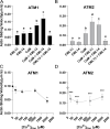
Actin sliding assays indicate that CaM, CML13, and CML14 can function as light chains for ATM1 and ATM2. (A) Actin sliding velocity of ATM1 in the presence of CaM, CML13, and CML14 alone or in different combinations of two putative MLCs. The concentrations used for CaM, CML13, and CML14 were 10, 30, and 30 µM, respectively. (B) Actin sliding velocity of ATM2 in the presence of CaM, CML13, and CML14 alone or in different combinations of two putative MLCs. The concentrations used for CaM, CML13, and CML14 were each 30 µM. (C) The Ca2+ sensitivity of ATM1 in the presence of CaM and CML13. The concentrations used for CaM and CML13 were 10 µM and 30 µM, respectively. (D) The Ca2+ sensitivity of ATM2 in the presence of CaM and CML13. The concentrations used for CaM and CML13 were 10 µM and 30 µM, respectively. Motility values are the mean ±SD and were determined by measuring the displacements of actin filaments that were smoothly moving for distances >10 μm as described in the Materials and methods.
Fig. 8.
Fig. 8.
Hypocotyl lengths of Col-0, myosin viii4KO, and Dex-inducible CML13,14 hpRNAi lines. Seeds were sown onto 0.5× MS agar plates with (treatment) or without (control) 5 µM Dex and cold stratified in the dark at 4 °C for 48 h. Plates were then moved to the growth chamber, still in the dark, and the hypocotyls were allowed to elongate for 5 d before imaging (A) and measuring the hypocotyl lengths with ImageJ (B). Boxes show the mean ±SD of each line on each of the control or treatment plates, whiskers portray the range of the data acquired, and each point is a single biological replicate (one-way ANOVA with Tukey’s test for multiple comparisons, with 20–45 biological replicates, *P<0.05, **P<0.01, ***P<0.001, ****P<0.0001). White bar in (A)=1 cm, and all images are of the same magnification.
Fig. 9.
Fig. 9.
Working model of CaM, CML13, and CML14 as light chains for Arabidopsis myosins ATM1 and ATM2. Actin-binding, catalytic myosin head domains are presented in red, the neck regions are decorated with light chains represented as blue (CaM), green (CML13/14), or gray (any of CaM/CML13/14) ovals, the coiled-coil dimerization regions are in yellow, and the cargo-binding tail domains are dark blue, three-quarter circles. Light chains bind to the IQ domains within the neck region of myosins to provide the leverage and structural integrity needed for the power stroke. We speculate that CaM is the preferred light chain at the IQ1 position (nearest the head) for both ATM1 and ATM2, whereas CML13 or CML14 are preferred light chains at the IQ2 position. Other IQ domains did not show a clear preference in our tests and may be occupied by CaM, CML13, CML14, or possibly other light chains.

Similar articles

Cited by

References

    1. Abu-Abied M, Belausov E, Hagay S, Peremyslov V, Dolja V, Sadot E. 2018. Myosin XI-K is involved in root organogenesis, polar auxin transport, and cell division. Journal of Experimental Botany 69, 2869–2881. - PMC - PubMed
    1. Abu-Abied M, Golomb L, Belausov E, Huang S, Geiger B, Kam Z, Staiger CJ, Sadot E. 2006. Identification of plant cytoskeleton-interacting proteins by screening for actin stress fiber association in mammalian fibroblasts. The Plant Journal 48, 367–379. - PubMed
    1. Alaimo A, Malo C, Areso P, Aloria K, Millet O, Villarroel A. 2013. The use of dansyl-calmodulin to study interactions with channels and other proteins. Methods in Molecular Biology 998, 217–231. - PubMed
    1. Altman D. 2013. Myosin: fundamental properties and structure. In: Roberts GCK, ed. Encyclopaedia of biophysics. Berlin, Heidelberg: Springer, 1664–1671.
    1. Avisar D, Abu-Abied M, Belausov E, Sadot E, Hawes C, Sparkes IA. 2009. A comparative study of the involvement of 17 Arabidopsis myosin family members on the motility of Golgi and other organelles. Plant Physiology 150, 700–709. - PMC - PubMed

Publication types

LinkOut - more resources