. 2024 Jan 27;15(1):812.
doi: 10.1038/s41467-024-45202-5.

The cryo-EM structure of homotetrameric attachment glycoprotein from langya henipavirus

Affiliations

The cryo-EM structure of homotetrameric attachment glycoprotein from langya henipavirus

Yingying Guo et al. Nat Commun. .

Abstract

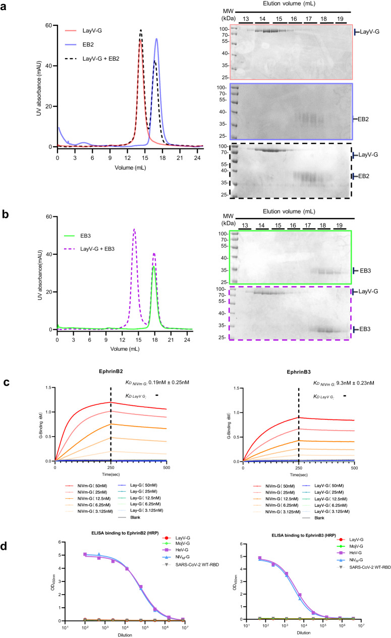
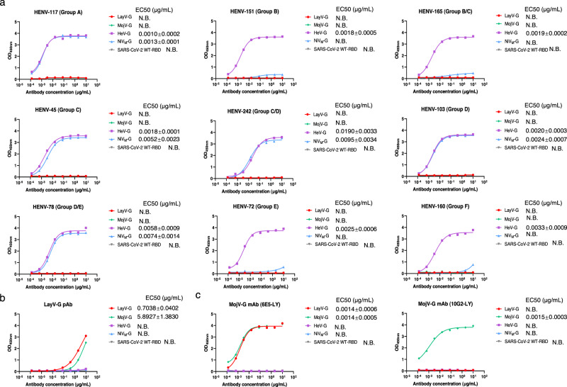
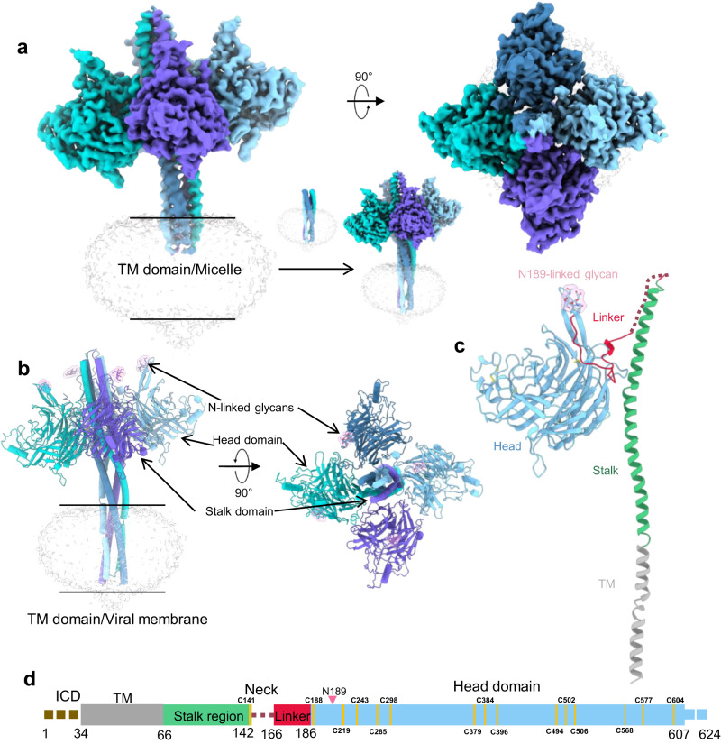
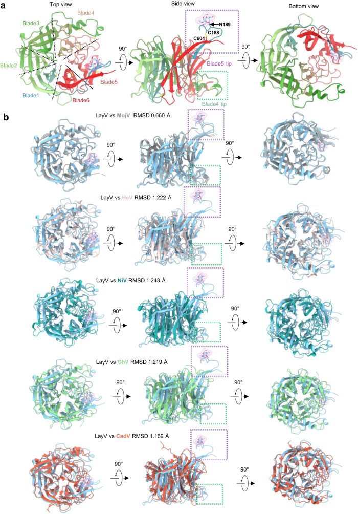
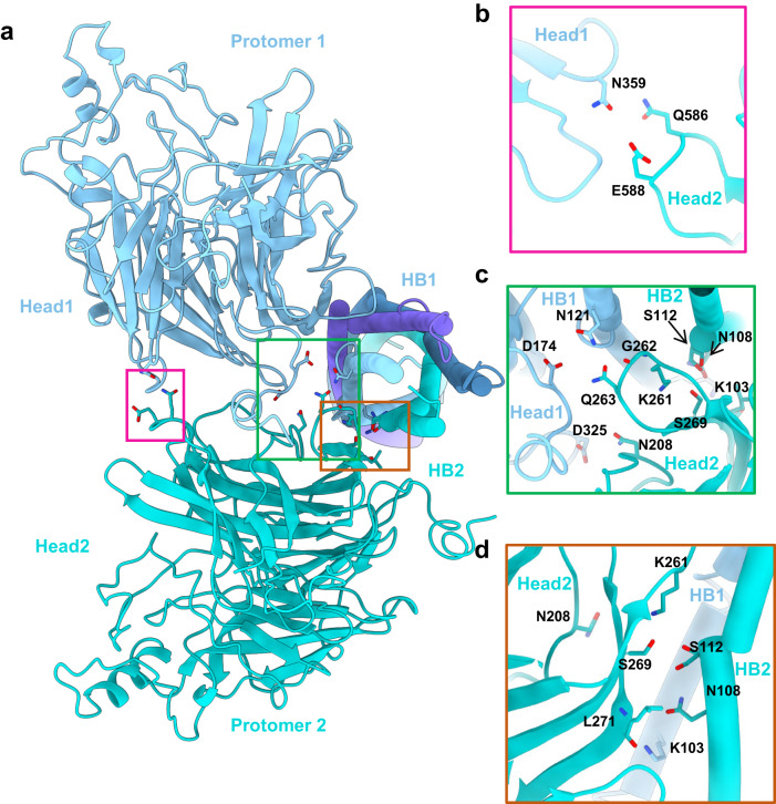
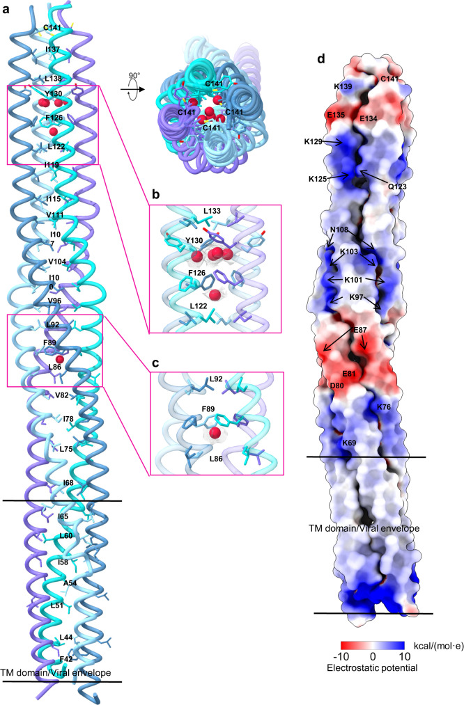
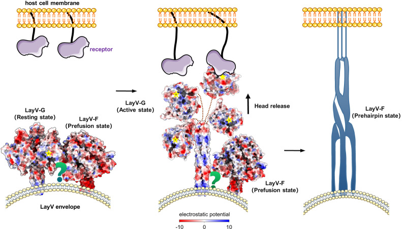
Langya Henipavirus (LayV) infection is an emerging zoonotic disease that has been causing respiratory symptoms in China since 2019. For virus entry, LayV's genome encodes the fusion protein F and the attachment glycoprotein G. However, the structural and functional information regarding LayV-G remains unclear. In this study, we revealed that LayV-G cannot bind to the receptors found in other HNVs, such as ephrin B2/B3, and it shows different antigenicity from HeV-G and NiV-G. Furthermore, we determined the near full-length structure of LayV-G, which displays a distinct mushroom-shaped configuration, distinguishing it from other attachment glycoproteins of HNV. The stalk and transmembrane regions resemble the stem and root of mushroom and four downward-tilted head domains as mushroom cap potentially interact with the F protein and influence membrane fusion process. Our findings enhance the understanding of emerging HNVs that cause human diseases through zoonotic transmission and provide implication for LayV related vaccine development.


Conflict of interest statement

The authors declare no competing interests.

Figures

Fig. 1
Fig. 1. Biochemical characterization of the LayV-G protein and binding affinity with Henipavirus receptors ephrinB2 and ephrinB3.
a, b Comparison of gel-filtration profile of LayV-G, human ephrinB2, ephrinB3. After LayV-G co-incubation with human ephrinB2/B3; LayV-G (Red) cannot form a complex with human ephrinB2 (Blue) or ephrinB3 (green) in gel-filtration (Shown on the left panel). The reducing-SDS-PAGE of fractions collected from gel-filtration (Shown on the right panel). c Bio-layer Interferometry (BLI) data of binding affinity of ephrinB2 (left) & ephrinB3 (right) and purified LayV-G and NiV-G. LayV-G (Blue scatter from dark to light) and NiV-G (Orange scatter from dark to light) were used as analytes in solution, with concentrations ranging from 50 nM to 3.125 nM. d ELISA binding of serious diluted soluble recombinant ephrinB2 and ephrinB3 conjugated with HRP to various HNV-G ectodomain (LayV-G (red), MojV-G (green), HeV-G (purple) and NiVM-G (blue)), SARS-CoV-2-WT (grey) served as a negative control. One representative curve of three independent experiments performed in technical duplicate are shown. EphrinB2-HRP and ephrinB3-HRP were separately serial diluted starting with 1:100 and 1:50. Source data are provided as a Source Data file.
Fig. 2
Fig. 2. The antigenicity of LayV-G protein in comparison to HeV-G, NiV-G and MojV-G proteins.
a Indirect ELISA detection of representative human anti-HeV-G mAbs from nine groups (A, B, B/C, C, C/D, D, D/E, E, F) binding to recombinant G proteins ectodomain of LayV-G (red), MojV-G (green), HeV-G (purple) and NiVM-G (blue). SARS-CoV-2 WT-RBD (grey) served as a non-reactive antigen for antibody targeting G protein of henipaviruses. b, c ELISA detection of rabbit derived anti-LayV-G polyclonal antibody and two MojV-G mAbs against soluble LayV-G, MojV-G, HeV-G, NiVM-G, and SARS-CoV-2 WT-RBD are shown. One representative curve of three independent experiments performed in technical duplicate are shown. The 50% effective concentration (EC50) values of tested antibodies are calculated on mean values of three independent experiments. Source data are provided as a Source Data file. Data are represented as mean ± SD. N.B. no binding.
Fig. 3
Fig. 3. Architecture of the nearly full-length LayV-G.
a The left panel shows the overall cryo-EM map of LayV-G (EMDB ID: EMD-36741), while the right panel displays top views of the overall structure. The structure consists of four homologous protomers, which are colored in blue, cyan, purple, and light blue, respectively. The low-resolution local refinement map of transmembrane was superimposed to the whole map. b The LayV-G model is represented in a cartoon diagram (PDB ID: 8JZB). The protomers are colored as described in a. The glycosylation site surface is highlighted in pink color. c In this panel, a single protomer is depicted. The head region is shown in light blue color, the glycosylation site surface in pink, the linker in red, the stalk in green, and the transmembrane (TM) domain in gray. d A linear representation of the full-length LayV-G is displayed. The intracellular domain (ICD) is colored in khaki, the transmembrane domain (TM) in gray. Unresolved density is indicated by a dashed line, while resolved density is represented by rectangles.
Fig. 4
Fig. 4. The feature of head domain of LayV-G and structure alignment with several HNVs.
a The left panel shows the top view from a vertical direction of the viral membrane (PDB ID:8JZB). The middle panel displays the side view, while the right panel depicts the bottom view of the head domain of LayV-G. The head domain is colored in a gradient from light blue, green, to red, representing the N-terminus to C-terminus orientation. Two special tips of blade4 and blade5 were boxed by a green and a purple dotted line boxed, respectively. Cysteine residues that form disulfide bonds in two tips are shown as sticks and labeled. b A structure alignment is performed with five typical HNVs attachment glycoprotein. The alignment includes MojV (colored in gray, PDB ID: 5NOP), HeV (colored in light pink, PDB ID: 2VSK), NiV (colored in deep green, PDB ID: 2VWD), GhV (colored in green, PDB ID: 4UF7), and CedV (colored in orange, PDB ID: 6P7Y).The green and purple dotted line boxes exhibit the special tips at the head domain of blade4 and blade5 in LayV-G are almost identical to those in MojV, and different from HeV, NiV, GhV and CedV.
Fig. 5
Fig. 5. The head and HB domain interaction.
a Two adjacent protomers (PDB ID:8JZB) are depicted, with one shown in blue color and the other in cyan color. This panel displays a detailed view of the interface between head1 from protomer1 and head2 from its adjacent protomer2. This view is from the top. bd Amino acid interaction details in the interface.
Fig. 6
Fig. 6. The 4HB domain features.
a Four HB domains (PDB ID:8JZB) are represented by a ribbon diagram, with each domain colored in blue, light blue, cyan, and purple, respectively. The residues of the hydrophobic core are shown as sticks, emphasizing their location within the domains. b, c Cryo-EM density corresponding to putative sodium ions (red color) at 0.22 level in ChimeraX 1.6.1. and the surrounding amino-acid residues. d Electrostatic surface of 4HB domain. Electrostatic potential value was estimated in ChimeraX 1.6.1 using coulombic calculation method.
Fig. 7
Fig. 7. Proposed model of LayV-G infecting host cell.
In the proposed model, the homotetrameric LayV-G (PDB:8JZB) could bind to the prefusion state of LayV-F (PDB ID: 8FEJ) in the resting state. Once the host receptor binding, the head domain of G protein might undergo a notable conformational change. Then the variegated patterns of electrostatic features on the surface of the 4HB appear to be responsible for triggering the conversion of the LayV-F from a prefusion to a pre-hairpin state and then a post-fusion state, thus triggering the membrane fusion between virus and host cells. Structure surfaces are colored by electrostatic potentials, which were estimated in ChimeraX 1.6.1 by coulombic calculation method.

Similar articles

Cited by

References

    1. Eaton BT, Broder CC, Middleton D, Wang LF. Hendra and Nipah viruses: different and dangerous. Nat. Rev. Microbiol. 2006;4:23–35. doi: 10.1038/nrmicro1323. - DOI - PMC - PubMed
    1. Field H, et al. The natural history of Hendra and Nipah viruses. Microbes. Infect. 2001;3:307–314. doi: 10.1016/S1286-4579(01)01384-3. - DOI - PubMed
    1. Weatherman S, Feldmann H, de Wit E. Transmission of henipaviruses. Curr. Opin. Virol. 2018;28:7–11. doi: 10.1016/j.coviro.2017.09.004. - DOI - PMC - PubMed
    1. Madera S, et al. Discovery and Genomic Characterization of a Novel Henipavirus, Angavokely Virus, from Fruit Bats in Madagascar. J. Virol. 2022;96:e0092122. doi: 10.1128/jvi.00921-22. - DOI - PMC - PubMed
    1. Zhang XA, et al. A Zoonotic Henipavirus in Febrile Patients in China. N. Engl J. Med. 2022;387:470–472. doi: 10.1056/NEJMc2202705. - DOI - PubMed

Supplementary concepts