. 2024 Jan 27;7(1):135.
doi: 10.1038/s42003-024-05778-6.

Defined microbial communities and their soluble products protect mice from Clostridioides difficile infection

Affiliations

Defined microbial communities and their soluble products protect mice from Clostridioides difficile infection

Katya Douchant et al. Commun Biol. .

Abstract

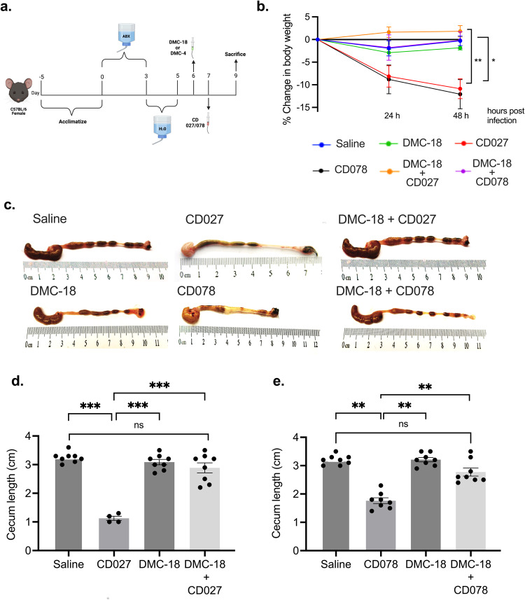
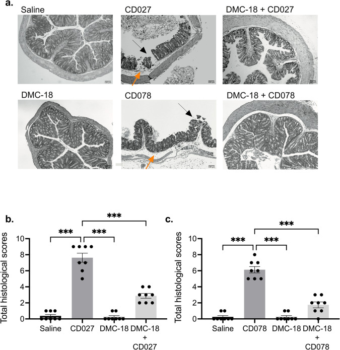
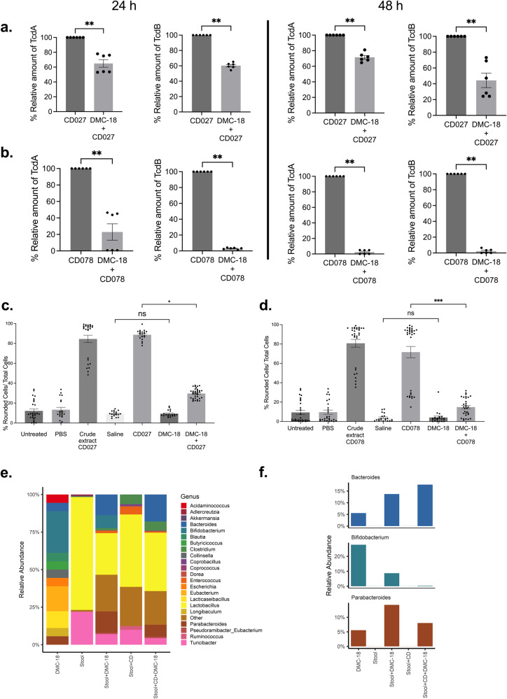
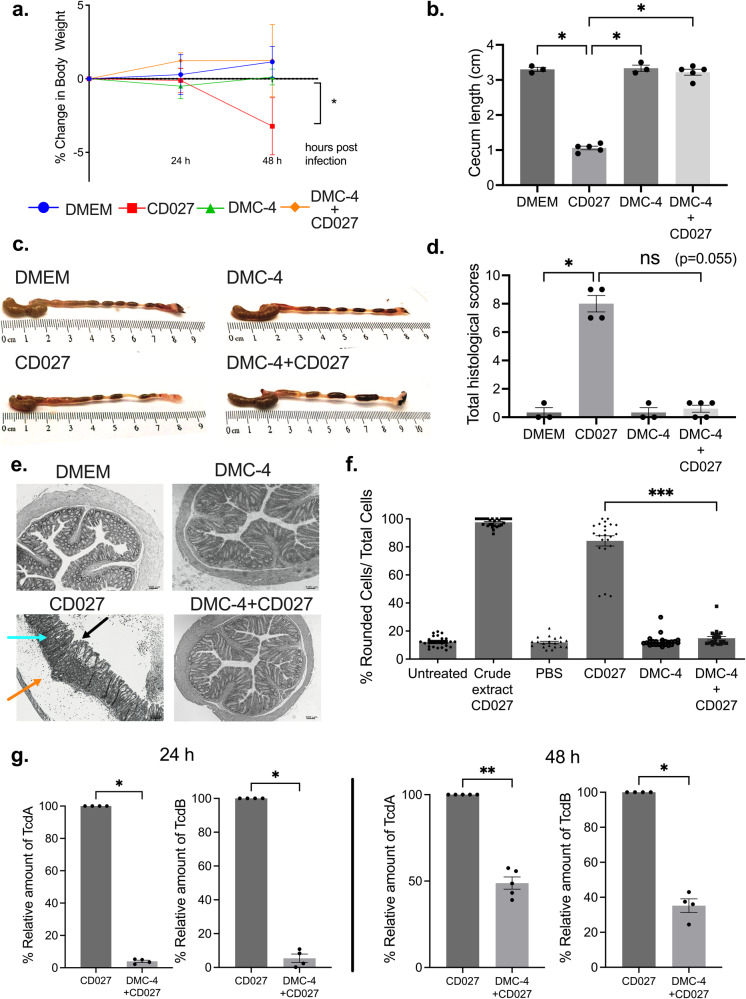
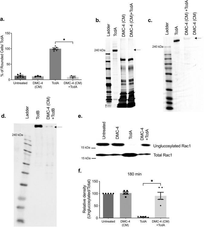
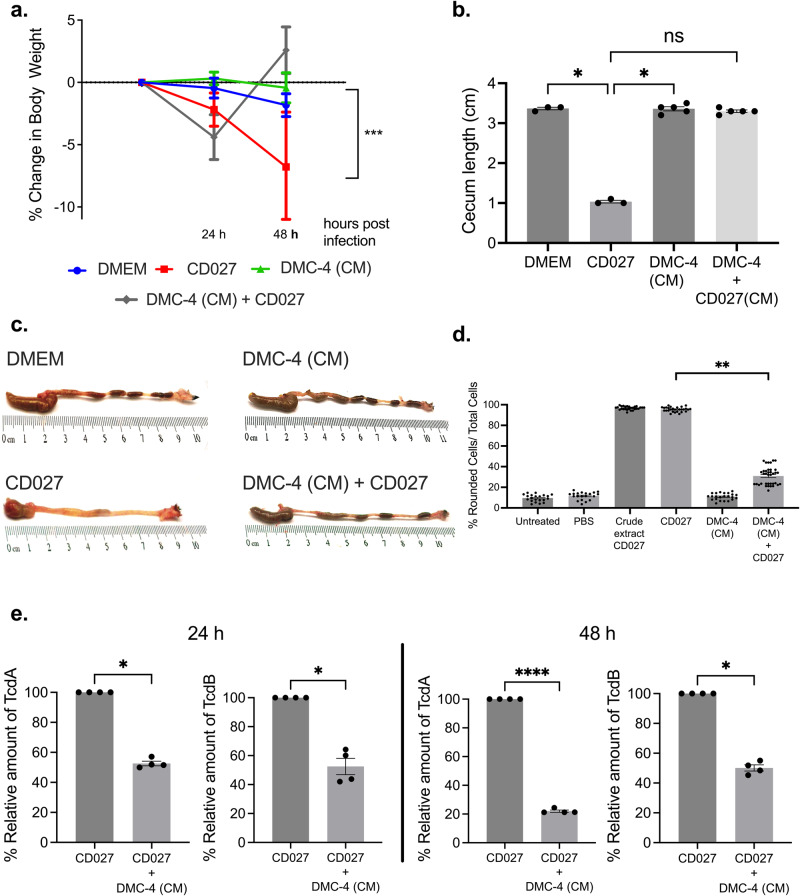
Clostridioides difficile is the leading cause of antibiotic-associated infectious diarrhea. The development of C.difficile infection is tied to perturbations of the bacterial community in the gastrointestinal tract, called the gastrointestinal microbiota. Repairing the gastrointestinal microbiota by introducing lab-designed bacterial communities, or defined microbial communities, has recently shown promise as therapeutics against C.difficile infection, however, the mechanisms of action of defined microbial communities remain unclear. Using an antibiotic- C.difficile mouse model, we report the ability of an 18-member community and a refined 4-member community to protect mice from two ribotypes of C.difficile (CD027, CD078; p < 0.05). Furthermore, bacteria-free supernatant delivered orally to mice from the 4-member community proteolyzed C.difficile toxins in vitro and protected mice from C.difficile infection in vivo (p < 0.05). This study demonstrates that bacteria-free supernatant is sufficient to protect mice from C.difficile; and could be further explored as a therapeutic strategy against C.difficile infection.


Conflict of interest statement

The authors declare no competing interests.

Figures

Fig. 1
Fig. 1. Pre-treatment with DMC-18 protected mice from CD-mediated weight loss and disease pathology.
a Schematic of the CD mouse model used in this study. Mice were given an antibiotic cocktail ad libitum in their drinking water for three days, and then water for the next two days. On day 6, mice were gavaged 150 μl of thawed DMC-18 mixtures (prepared in the proportions described in Table 1), followed by CD027 or CD078 challenge (1 × 105 CFU) on day 7. Mice were weighed, and their stools were collected daily. On day 9, mice were euthanatized by cervical dislocation. Blood, stool, colon, and cecum were harvested. Created with BioRender.com. b DMC-18 treated mice were protected from significant weight loss following CD027 or CD078 infection (two-way ANOVA with Tukey’s test, n = 8 for each group, *p < 0.05, **p < 0.01). c Representative images of the gross morphology of the ceca and colons from all groups of mice taken at the same magnification (20X). Uninfected mice and those exposed to DMC-18 prior to CDI had large ceca filled with feces and well-defined fecal pellets in the colon, while infected animals showed a reduction in their ceca size and demonstrated colitis. d Ceca were measured to compare sizes between uninfected, CD-infected and CD-infected+DMC-18 mice with CD027 and e CD078. Mice infected with CD had significant colonic shortening that was prevented when given DMC-18. A one-way ANOVA with Dunnett’s correction, n = 8 for each group was performed (***p < 0.001). Error bars represent standard error of the mean.
Fig. 2
Fig. 2. Histology of the murine colon revealed damage in CD-infected mice.
a Representative H&E stained images of the colon of all mouse groups. Mice treated with CD027 and CD078 had evidence of increased epithelial injury (black arrow), mucosal edema (orange arrow) and neutrophil infiltration. Scale bar, 100 μm. Histological scores by two individuals in a blinded fashion performed on all groups enumerating the degree of edema, neutrophilic infiltration, and epithelial cell damage following CD027 (b) and CD078 infection (c). The scores ranged from 0 (no damage) to 3 (severe damage/fulminant disease), for a total of 9. DMC-18 exposure prior to CD infection reduced CD-mediated damage in mice infected with CD027 or CD078. CD infected mice had significantly higher scores compared to CD + DMC-18 mice (Kruskal–Wallis with Dunn’s test, n = 8 for each group, ***p < 0.001). Error bars represent standard error of the mean.
Fig. 3
Fig. 3. DMC-18 pre-treatment reduced toxin levels in mouse stool.
CD + DMC-18 mice had significant reduction in relative concentrations of TcdA and TcdB in both CD027 (a) and CD078 (b) infected mice at 24 h and 48 h of infection (Mann–Whitney and a two-tailed test, n = 8 per group, **p < 0.01 for all groups tested in triplicate). Fibroblast cell rounding assays revealed that CD + DMC-18 mice had reduced toxin-mediated cytotoxicity in their fecal pellets compared to CD-mice infected with both CD027 c and CD078 d at 48 h post infection (Kruskal–Wallis with Dunn’s test was used to analyze the data, n = 8, **p < 0.01, ***p < 0.001). e 16S rRNA sequencing of murine stool from all groups demonstrated that DMC-18 led to a significant change in the relative abundance of bacteria in stool. f When comparing CD-infected mice treated with DMC-18 (Stool + CD + DMC-18) to CD-infected mice not exposed to DMC-18 we identified three bacterial genera only present in DMC-18, CD + DMC-18 but not CD-infected mice. Bifidobacterium, Parabacteroides, and Bacteroides were detected 24 h post-infection. These genera were absent prior to DMC-18 inoculation (stool) and in mice infected with CD but not treated with DMC-18 (Stool + CD). Operational taxonomic units were based on 97% identity; relative abundance is displayed. Error bars represent standard error of the mean.
Fig. 4
Fig. 4. Pre-treatment with DMC-4 protected mice from CD-mediated weight loss, disease pathology and reduced toxin levels in stool.
a Following the discovery that 4-members of DMC- 18 were found in the stool of mice protected from CDI, but not in the stool of CD-infected mice, a bacterial community of 4-bacteria (called DMC-4) was developed. DMC-4 treated mice were protected from significant weight loss following CD027 (two-way ANOVA with Tukey’s test, control animals n = 3 and infected animals n = 5, *p < 0.05). b Ceca from all mice were measured to compare sizes between uninfected, CD027-infected and DMC-4 + CD027 mice, similar to DMC-18; DMC-4 exposed mice were protected from ceca-shortening (one-way ANOVA with Dunnett’s correction, control animals n = 3 and infected animals n = 5, *p < 0.05, **p < 0.01). c Representative images of the gross morphology of the cecum and colon from all groups of mice taken at the same magnification (20X). d Histological scores of all treatment groups based on the degree of edema, neutrophilic infiltration, and epithelial cell damage following CD027. DMC-4 exposure prior to CD infection reduced CD-mediated damage in mice infected with CD027 (Kruskal–Wallis with Dunn’s test, control animals n = 3 and infected animals n = 5, *p < 0.05). e DMC-4 exposure reduces toxin activity and cytotoxicity of fecal pellets from CD027-infected mice Representative H&E stained images of the colon of all mouse groups demonstrated that CD027infected mice had evidence of increased epithelial injury (black arrow), mucosal edema (orange arrow) and neutrophil infiltration (blue arrow). Scale bar, 100 μm. f Cell rounding assays also demonstrated significantly lower toxin activity in stool from mice infected with CD027 + DMC-4 infected mice 48 h after the infection compared to the CD027 group (Kruskal-Wallis with Dunn’s test, ***p < 0.001). Pooled supernatants from stool pellets, control animals (n = 3) and infected animals (n = 5). Error bars represent standard error of the mean. g TcdA and TcdB quantifications in DMC-4-treated mice 24 and 48 h after CD027 infection were significantly lower than in CD-infected mice (t-Test with Mann–Whitney and two-tailed test, *p < 0.05. All groups were replicated in quadruplicate).
Fig. 5
Fig. 5. DMC-4 (CM) proteolyzed TcdA and TcdB and prevented glucosylation of Rac1.
a In addition to DMC-4 inhibiting toxin activity; conditioned media (CM) from DMC-4 also inhibited TcdA-mediated cell rounding in vitro (pooled results of assays run in triplicate, Kruskal-Wallis with Dunn’s test, p < 0.05). Silver staining (b) and western blot demonstrated the loss of purified TcdA (100 ng) (c) and TcdB (100 ng) (d) when incubated with DMC-4 (CM) for 1h. e Densitometry analysis was conducted to quantify glucosylation activity of Rac1 using DMC-4 (CM). Toxin-mediated glucosylation was inhibited (e, f) in cells treated with DMC-4 (CM) supernatants (p < 0,05, One-way ANOVA with Dunnett’s test. Error bars represent standard error of the mean).
Fig. 6
Fig. 6. Pre-treatment with DMC-4 (CM) protected mice from CDI.
a DMC-4 (CM) treated mice were protected from significant weight loss following CD027 infection compared to CD-infected mice (two-way ANOVA with Tukey’s test, DMEM n = 3, CD027 n = 4, DMC-4 (CM) n = 4 and DMC-4 (CM) + CD027 n = 4, *p < 0.05). b DMC-4 CM + CD mice were protected from ceca shortening compared to CD-infected mice (one-way ANOVA with Dunnett’s correction, DMEM n = 3, CD027 n = 4, DMC-4 (CM) n = 4 and DMC-4 (CM) + CD027 n = 4, *p < 0.05). c Representative images of the gross morphology of the ceca and colons from all groups of mice taken at the same magnification (×20) demonstrated that uninfected mice and those exposed to DMC-4 (CM) prior to CD infection had large ceca filled with feces and fecal pellets in the colon, while CD infected mice had reduced cecum size and morphological evidence of colitis. d Stool from mice infected with CD027 resulted in significantly higher toxin-mediated cell rounding compared to DMC-4 CM + CD027 (Kruskal-Wallis with Dunn’s test, **p < 0.01). Pooled supernatants from stool pellets, control animals n = 3 and infected animals n = 5. e TcdA and TcdB quantifications in DMC-4 (CM) treated mice after CD027 infection demonstrated that CD + DMC-4 CM mice had significantly lower levels of TcdA and TcdB at both 24 h and 48 h post-infection (Mann–Whitney test was used to analyze the data (*p < 0.05). All groups were carried out in triplicate). Error bars represent standard error of the mean.

Similar articles

Cited by

References

    1. Round JL, O’Connell RM, Mazmanian SK. Coordination of tolerogenic immune responses by the commensal microbiota. J. Autoimmun. 2010;34:J220–J225. doi: 10.1016/j.jaut.2009.11.007. - DOI - PMC - PubMed
    1. Mazmanian SK, Liu CH, Tzianabos AO, Kasper DL. An immunomodulatory molecule of symbiotic bacteria directs maturation of the host immune system. Cell. 2005;122:107–118. doi: 10.1016/j.cell.2005.05.007. - DOI - PubMed
    1. Kamada N, Seo S-U, Chen GY, Nunez G. Role of the gut microbiota in immunity and inflammatory disease. Nat. Rev. Immunol. 2013;13:321–335. doi: 10.1038/nri3430. - DOI - PubMed
    1. Pickard JM, Nunez G. Pathogen colonization resistance in the gut and its manipulation for improved health. Am. J. Pathol. 2019;189:1300–1310. doi: 10.1016/j.ajpath.2019.03.003. - DOI - PMC - PubMed
    1. Cleary RK. Clostridium difficile-associated diarrhea and colitis: clinical manifestations, diagnosis, and treatment. Dis. Colon Rectum. 1998;41:1435–1449. doi: 10.1007/BF02237064. - DOI - PubMed

Publication types

Substances