. 2024 Feb:45:103983.
doi: 10.1016/j.pdpdt.2024.103983. Epub 2024 Jan 27.

Combination of vitamin D and photodynamic therapy enhances immune responses in murine models of squamous cell skin cancer

Affiliations

Combination of vitamin D and photodynamic therapy enhances immune responses in murine models of squamous cell skin cancer

Sanjay Anand et al. Photodiagnosis Photodyn Ther. 2024 Feb.

Abstract

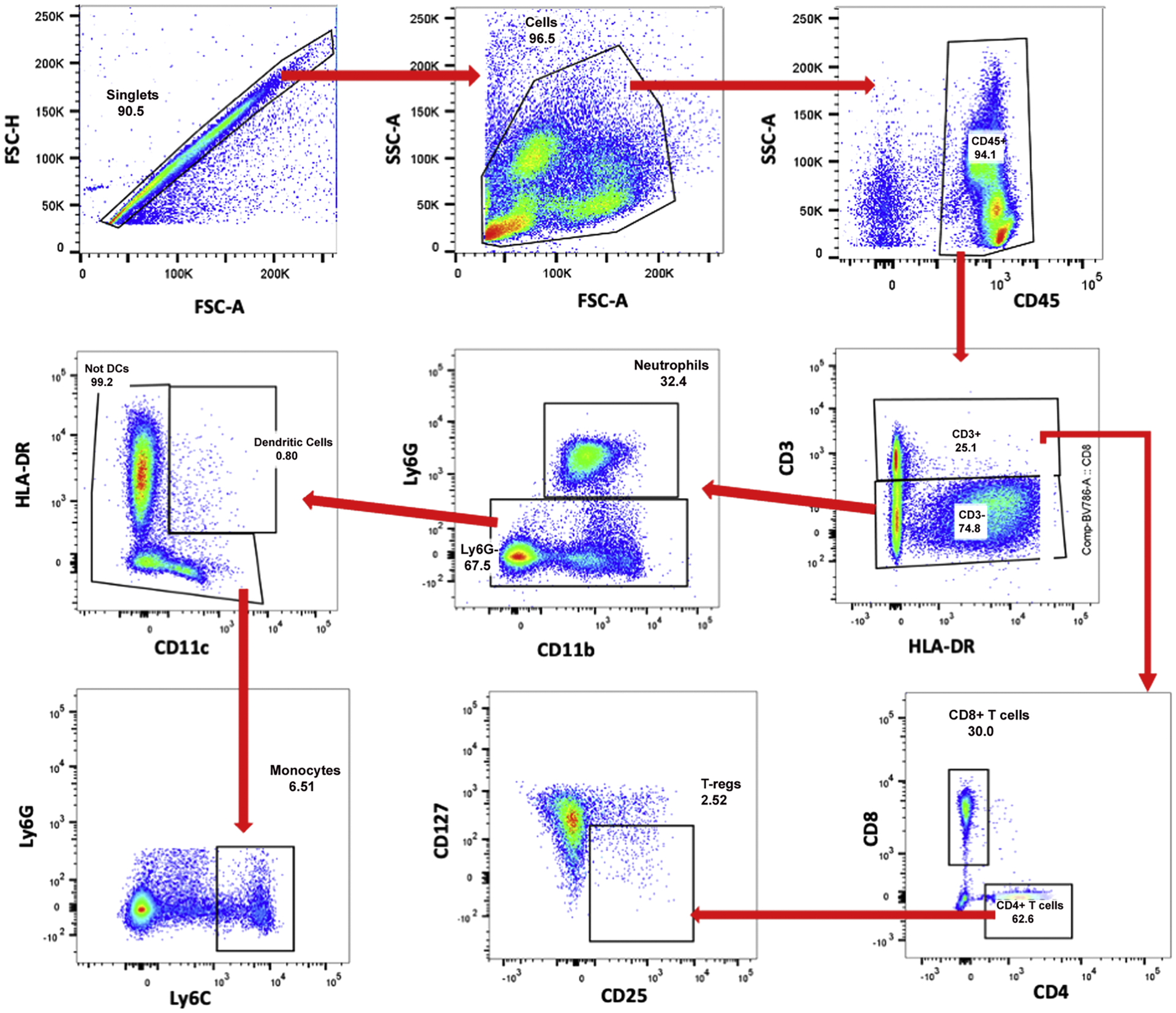
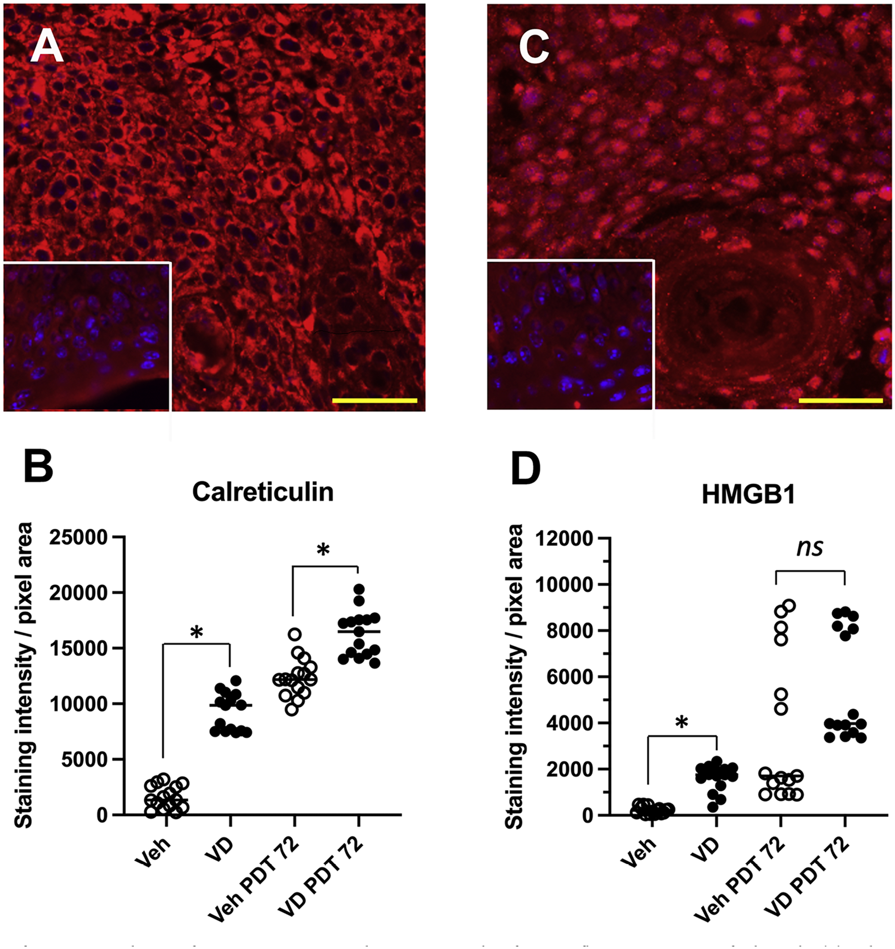
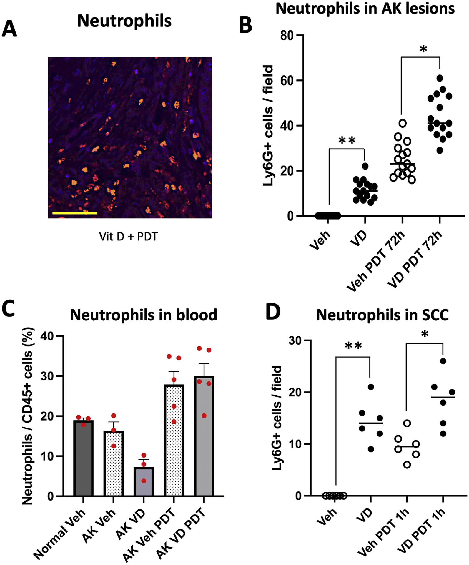
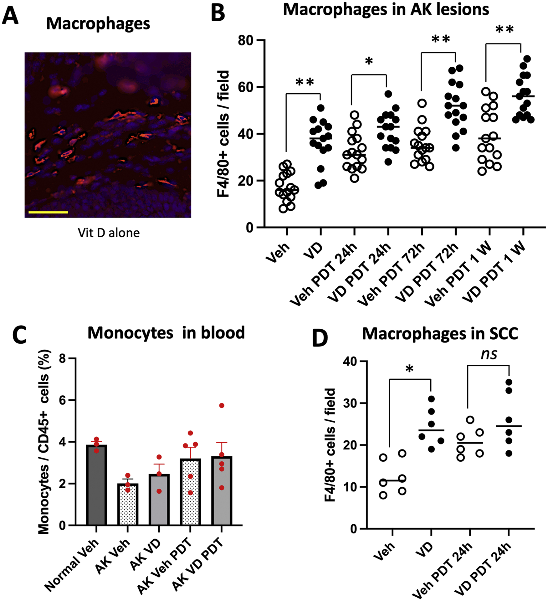
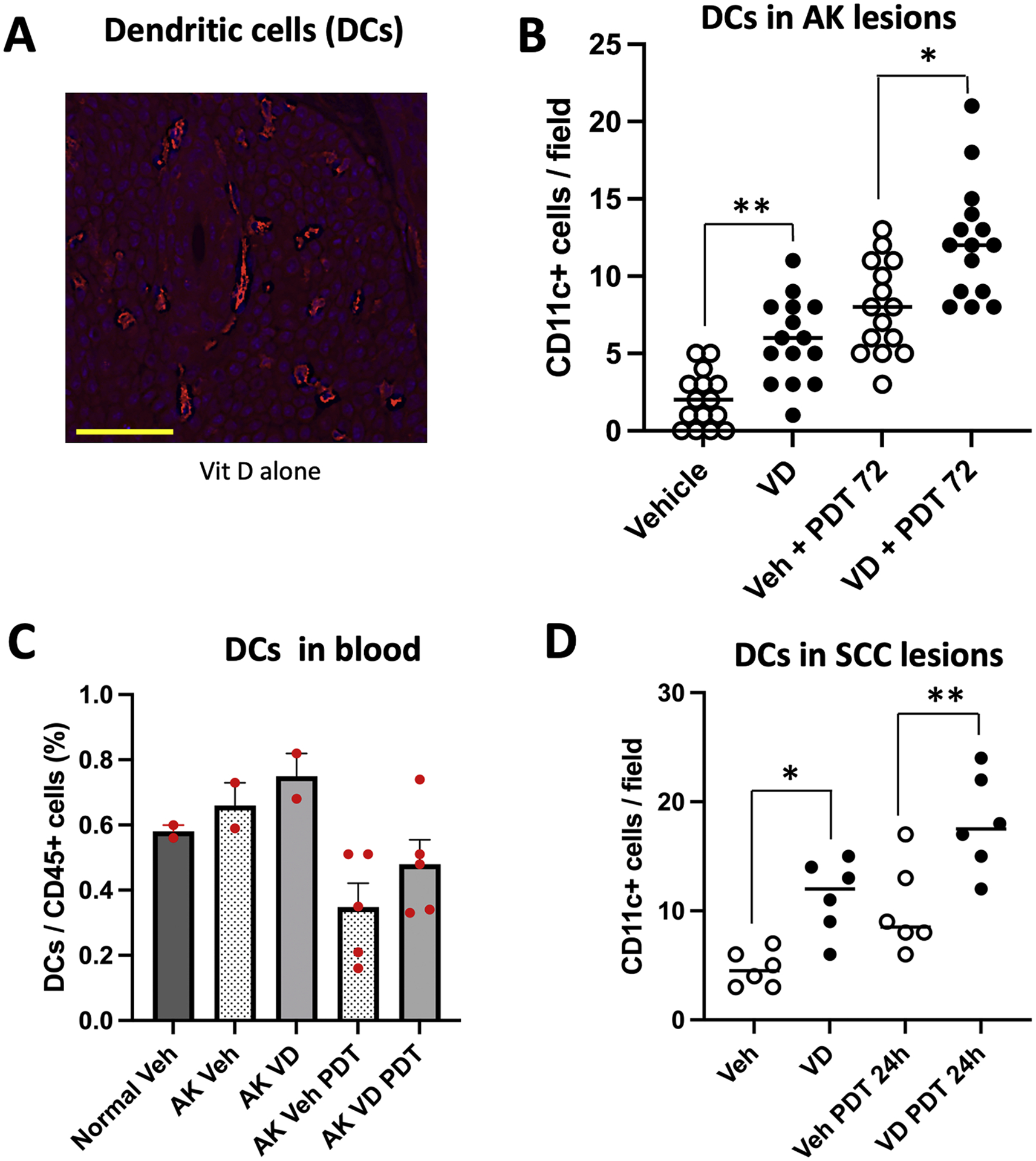
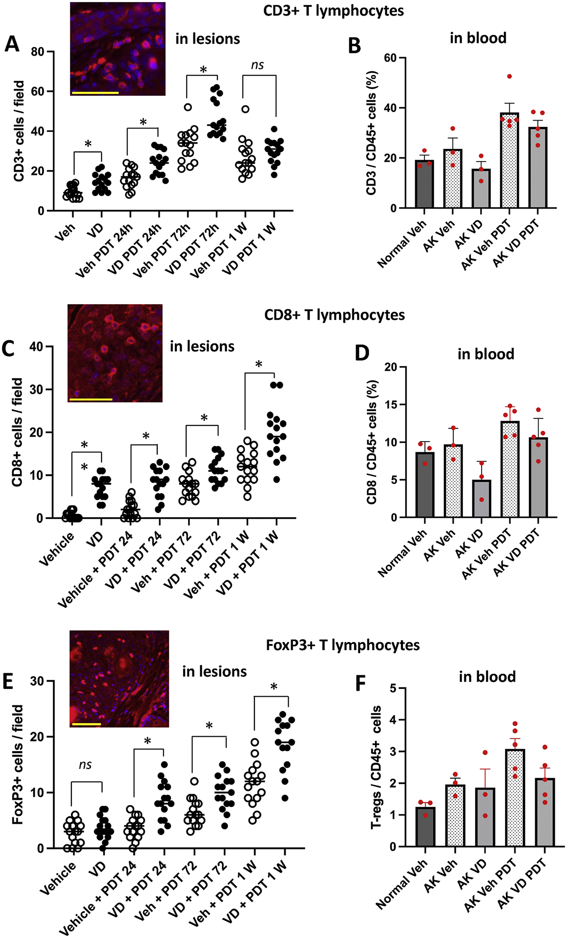
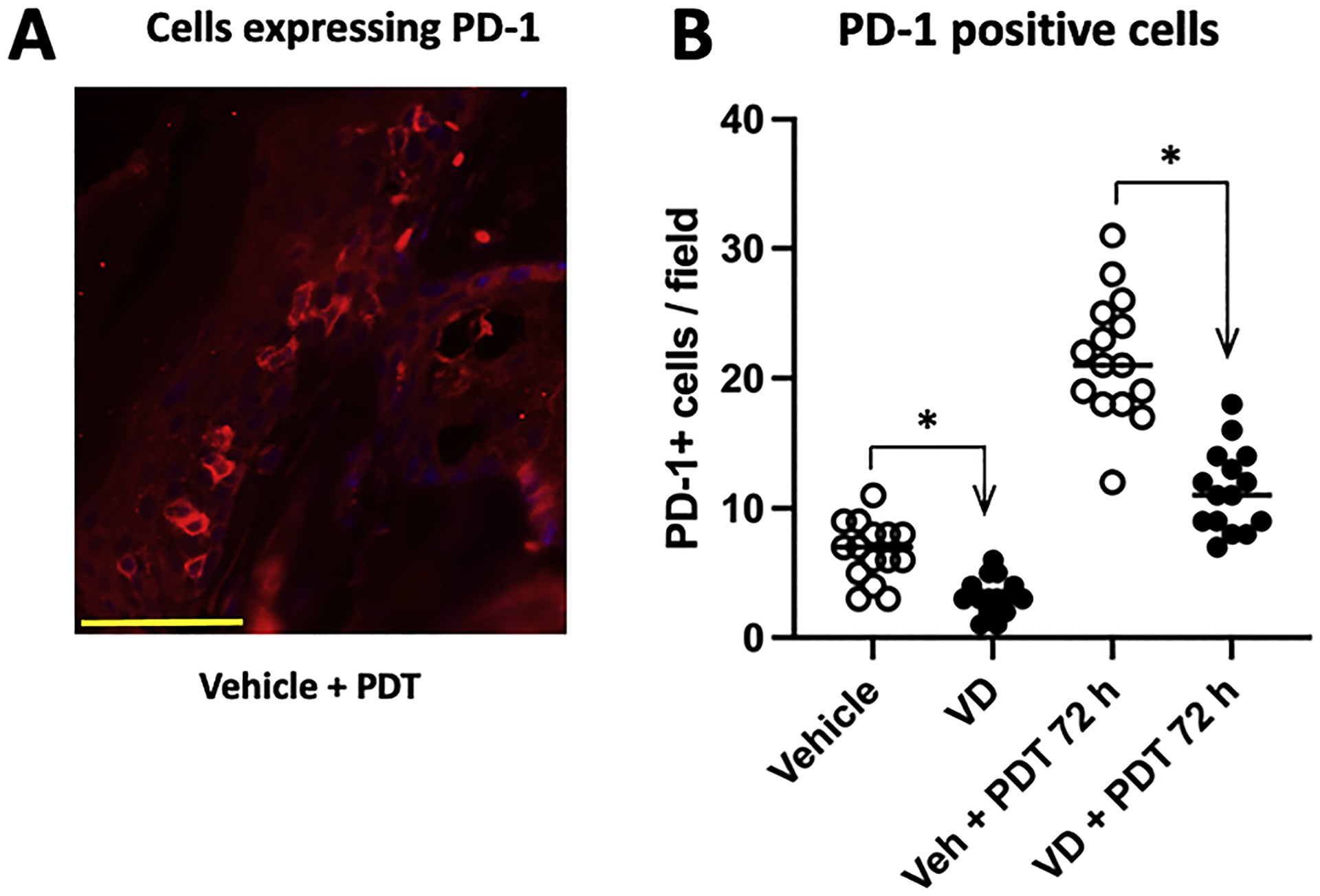
Improved treatment outcomes for non-melanoma skin cancers can be achieved if Vitamin D (Vit D) is used as a neoadjuvant prior to photodynamic therapy (PDT). However, the mechanisms for this effect are unclear. Vit D elevates protoporphyrin (PpIX) levels within tumor cells, but also exerts immune-modulatory effects. Here, two murine models, UVB-induced actinic keratoses (AK) and human squamous cell carcinoma (A431) xenografts, were used to analyze the time course of local and systemic immune responses after PDT ± Vit D. Fluorescence immunohistochemistry of tissues and flow analysis (FACS) of blood were employed. In tissue, damage-associated molecular patterns (DAMPs) were increased, and infiltration of neutrophils (Ly6G+), macrophages (F4/80+), and dendritic cells (CD11c+) were observed. In most cases, Vit D alone or PDT alone increased cell recruitment, but Vit D + PDT showed even greater recruitment effects. Similarly for T cells, increased infiltration of total (CD3+), cytotoxic (CD8+) and regulatory (FoxP3+) T-cells was observed after Vit D or PDT, but the increase was even greater with the combination. FACS analysis revealed a variety of interesting changes in circulating immune cell levels. In particular, neutrophils decreased in the blood after Vit D, consistent with migration of neutrophils into AK lesions. Levels of cells expressing the PD-1+ checkpoint receptor were reduced in AKs following Vit D, potentially counteracting PD-1+ elevations seen after PDT alone. In summary, Vit D and ALA-PDT, two treatments with individual immunogenic effects, may be advantageous in combination to improve treatment efficacy and management of AK in the dermatology clinic.

Keywords: Immune response; Murine model; Non-melanoma skin cancer; Photodynamic therapy; Vitamin D.


Conflict of interest statement

Declarations of competing interest None.

Figures

Fig. 1.
Fig. 1.
Gating strategy to identify and quantify immunologic cell types from flow cytometry data. Each blood sample was analyzed using the same gating strategy. First, single cells were gated using FSC-A and FSC–H (forward scattering), followed by total cells using FSC-A and SSC-A (side scattering). From total cells, total immune cells (CD45+) were gated. Following this, total T cells (CD3+) and non-T cells (CD3−) were gated. All CD3+ T cells were gated to discriminate CD8 T cells (CD8+) and CD4 T cells (CD4+). From CD4+ T cells, T-regs (CD25+, CD127−) were gated. From CD3− non-T cells, neutrophils (Ly6G+, CD11b+) were gated. From Ly6G− cells, dendritic cells (HLA-DR+, CD11c+) cells were gated. Finally, monocytes (Ly6C+) were gated from non-dendritic cells.
Fig. 2.
Fig. 2.
Expression of DAMPS in AK lesions after exposure to Vit D and/or PDT. Examples of immunofluorescent staining of calreticulin (A) and HMGB1 (C), at 72 h after Vit D pretreatment and PDT exposure. Insets, staining controls with no primary antibody. (B, D) Quantification of staining intensity for calreticulin and HMGB1, respectively, from 5 mice (3 lesions/mouse) per experimental condition. Statistical comparisons: Mann-Whitney non-parametric tests, (*), p < 0.0001; ns, not significant. Scale bars, 50 μM.
Fig. 3.
Fig. 3.
Effects of Vit D and PDT on recruitment of neutrophils into AK and SCC lesions. (A) Examples of immunofluorescent staining (IF) of neutrophils and (B) quantification of numbers of neutrophils per high-power field, in AK lesions. Mice were treated as follows: Veh, vehicle alone. VD, topical Vitamin D pretreatment alone. Veh PDT 72 h, lesions harvested 72 h after PDT alone. VD PDT 72 h, lesions harvested 72 h after PDT combined with Vit D pretreatment. Data shown are from 5 mice (3 images/lesion/mouse) per experimental condition. Differences are significant by nonparametric tests, namely, the Wilcoxon signed-rank test (**, p < 0.0001) and the Mann-Whitney test (*, p< 0.0001). (C) FACS analysis of neutrophils in peripheral blood from the mice with AK, collected 72 h after PDT. Three mice each for normal SKH-1 and SKH-1 mice with vehicle-treated AKs, and 5 mice each for Vit D, PDT and Vit d-PDT treatment conditions were analyzed. Note that differences among different treatment groups were statistically not significant. (D) Neutrophils in subcutaneous A431 SCC tumors harvested 1 hour after PDT. Data shown represent 3 mice (2 tumors/mouse) per experimental condition. Differences are significant, Wilcoxon signed-rank test, (**, p < 0.05), or Mann-Whitney test (*, p< 0.01). Scale bar, 50 μM.
Fig. 4.
Fig. 4.
Effects of VD and PDT on recruitment of macrophages into AK and SCC lesions. (A) Representative images, and (B) quantification of macrophages (F4/80 positive cells) in AK lesional tissue, harvested at time zero or at 24 h, 72 h, or 1 week after PDT, with or without Vitamin D pretreatment. Data shown are from 5 mice (3 images/lesion/mouse) per experimental condition. Differences are significant by Mann-Whitney test (*, p<0.005; **, p< 0.0001). (C) Monocytes in peripheral blood from AK-bearing mice at 72 h after PDT; see Fig. 1 for gating scheme used to identify monocytes. Three mice each for normal SKH-1 and SKH-1 mice with vehicle-treated AKs, and 5 mice each for Vit D, PDT and Vit d-PDT treatment conditions were analyzed. (D) Macrophages in subcutaneous A431 SCC tumor, harvested 24 h after PDT. Data shown represent 3 mice (2 tumors/mouse) per experimental condition. Significant difference, Mann-Whitney test (*, p< 0.005); ns, not significant. Scale bar, 50 μM.
Fig. 5.
Fig. 5.
Effects of VD and PDT on recruitment of dendritic cells into AK and SCC lesions. (A) Representative images, and (B) quantification of dendritic cells in AK lesions, either before or 72 h after PDT. Data shown are from 5 mice (3 images/lesion/mouse) per experimental condition. Differences are significant by Mann-Whitney test. (* p< 0.002; ** p<0.0001). (C) Dendritic cells in blood, at 72 h post PDT. Three mice each for normal SKH-1 and SKH-1 mice with vehicle-treated AKs, and 5 mice each for Vit D, PDT and Vit d-PDT treatment conditions were analyzed. (D) Dendritic cells in subcutaneous A431 SCC tumors, at 24 h post PDT. Data shown represent 3 mice (2 tumors/mouse) per experimental condition. Differences are significant by Mann-Whitney test. (* p< 0.01; ** p<0.005). Scale bar, 50 μM.
Fig. 6.
Fig. 6.
Effects of VD and PDT on recruitment of T cells into AK lesions at specified times after PDT (A, C, E), and in blood at 72 h after PDT (B, D, F). Data shown for immunofluorescence are from 5 mice (3 images/lesion/mouse) per experimental condition. For FACS analysis, three mice each for normal SKH-1 and SKH-1 mice with vehicle-treated AKs, and 5 mice each for Vit D, PDT and Vit d-PDT treatment conditions were analyzed. Insets, representative images of T-cells staining in AK lesional tissue. Changes in cell numbers are statistically different for the following comparisons, by Mann-Whitney test. (A) * p< 0.001; (B) * p< 0.005; ** p<0.0001; (C) * p< 0.005. Scale bars, 50 μM.
Fig. 7.
Fig. 7.
Number of cells expressing PD-1 in AK lesions. AK-bearing mice were either untreated or treated with Vit D alone, PDT alone, or combined Vit d-plus-PDT; lesions were biopsied 72 h post-PDT and the number of cells expressing checkpoint inhibitor receptor PD-1 were scored by IF in AK tissue sections (A). Whereas PDT increased the number of PD-1+ cells in lesions, Vit D (either alone or in combination with PDT) led to a reduction in the number of these cells (B). Data shown for immunofluorescence are from 5 mice (3 images/lesion/mouse) per experimental condition. Differences are significant by Mann-Whitney test, * p < 0.0001. Scale bar, 50 μM.

Similar articles

Cited by

References

    1. Rogers HW, Weinstock MA, Feldman SR, Coldiron BM, Incidence estimate of nonmelanoma skin cancer (keratinocyte carcinomas) in the U.S. population, 2012, JAMA Dermatol. 151 (10) (2015) 1081–1086. - PubMed
    1. Siegel RL, Miller KD, Wagle NS, Jemal A, Cancer statistics, 2023, CA Cancer J. Clin 73 (1) (2023) 17–48. - PubMed
    1. Ziegler A, Jonason AS, Leffell DJ, Simon JA, Sharma HW, Kimmelman J, Remington L, Jacks T, Brash DE, Sunburn and p53 in the onset of skin cancer, Nature 372 (6508) (1994) 773–776. - PubMed
    1. Brash DE, Sunlight and the onset of skin cancer, Trends Genet. 13 (10) (1997) 410–414. TIG. - PubMed
    1. Bowden GT, Prevention of non-melanoma skin cancer by targeting ultraviolet-B-light signalling, Nat. Rev. Cancer 4 (1) (2004) 23–35. - PubMed

MeSH terms