Review
. 2024 Jan 28;22(1):41.
doi: 10.1186/s12951-024-02298-7.

Exosome-based delivery strategies for tumor therapy: an update on modification, loading, and clinical application

Affiliations
Review

Exosome-based delivery strategies for tumor therapy: an update on modification, loading, and clinical application

Qian Yang et al. J Nanobiotechnology. .

Abstract

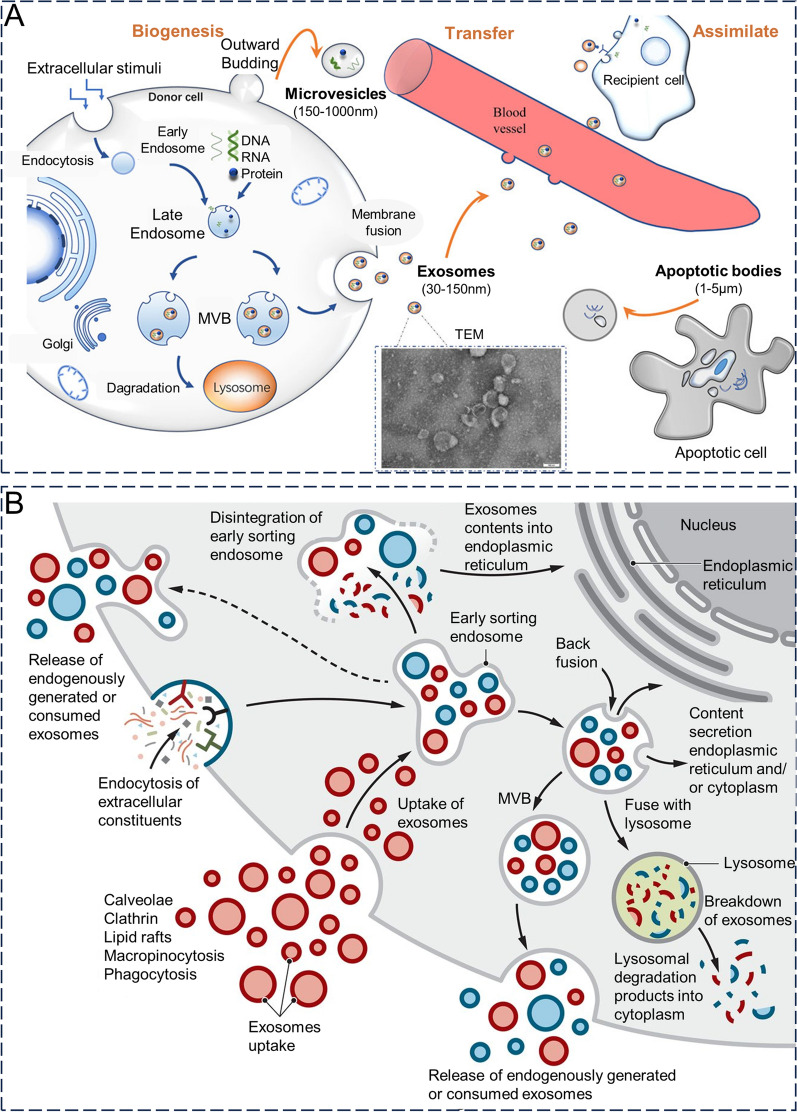
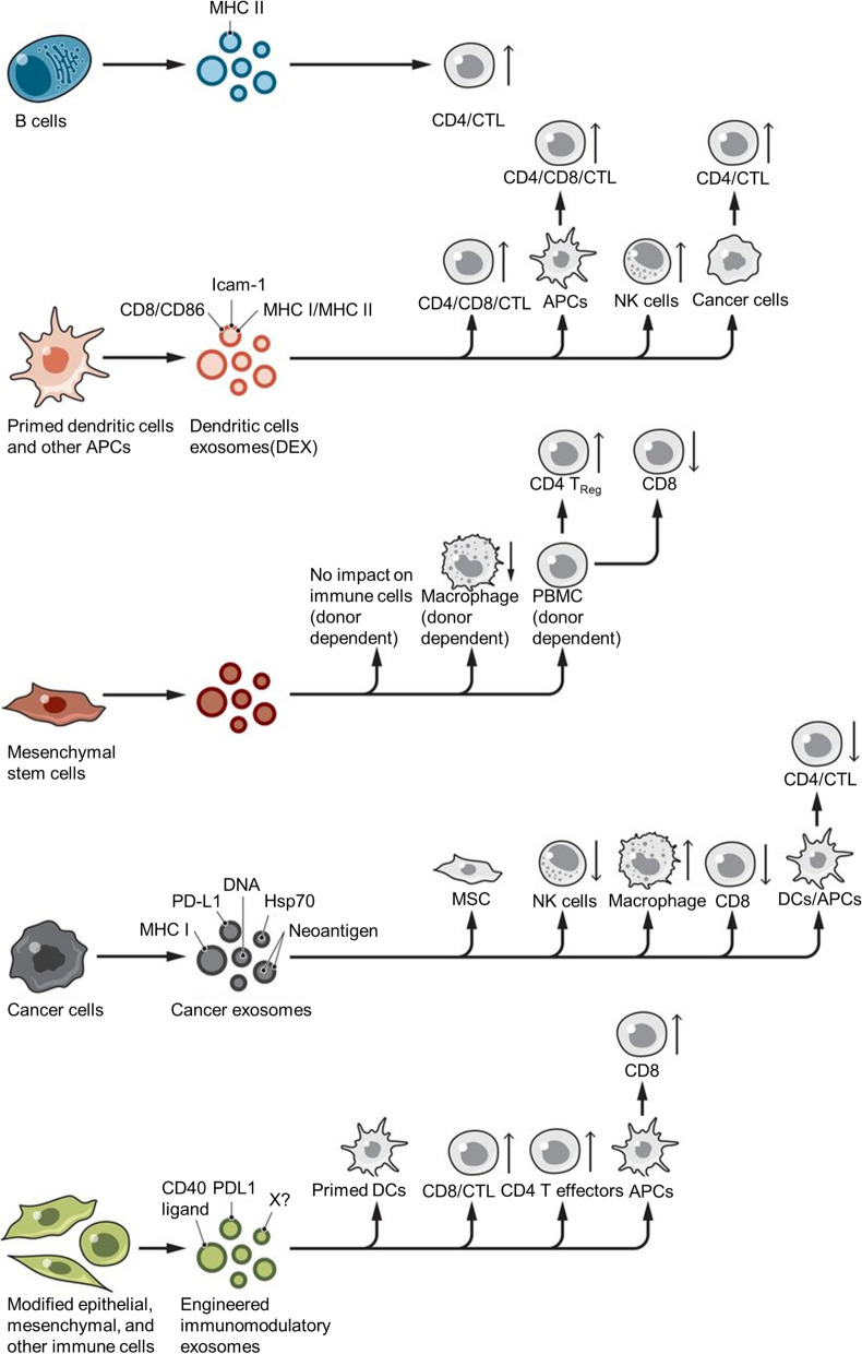
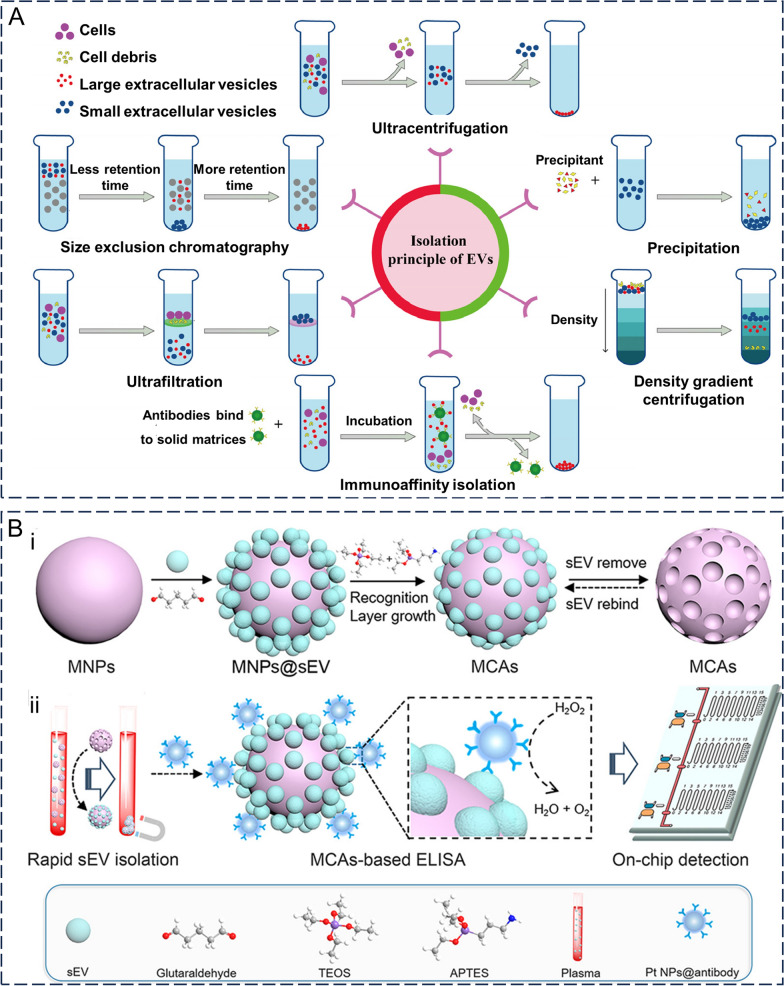
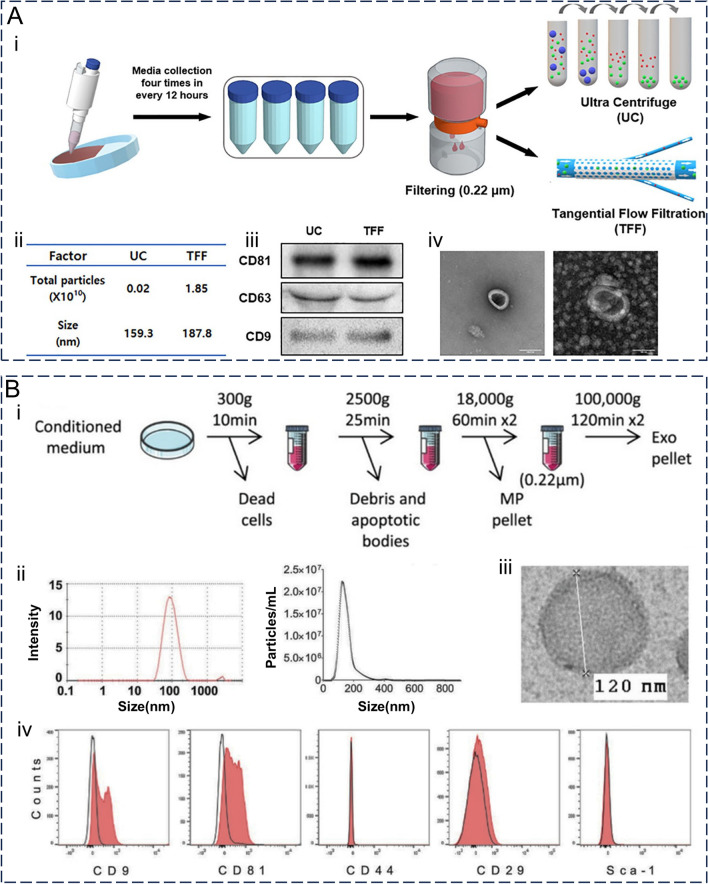
Malignancy is a major public health problem and among the leading lethal diseases worldwide. Although the current tumor treatment methods have therapeutic effect to a certain extent, they still have some shortcomings such as poor water solubility, short half-life, local and systemic toxicity. Therefore, how to deliver therapeutic agent so as to realize safe and effective anti-tumor therapy become a problem urgently to be solved in this field. As a medium of information exchange and material transport between cells, exosomes are considered to be a promising drug delivery carrier due to their nano-size, good biocompatibility, natural targeting, and easy modification. In this review, we summarize recent advances in the isolation, identification, drug loading, and modification of exosomes as drug carriers for tumor therapy alongside their application in tumor therapy. Basic knowledge of exosomes, such as their biogenesis, sources, and characterization methods, is also introduced herein. In addition, challenges related to the use of exosomes as drug delivery vehicles are discussed, along with future trends. This review provides a scientific basis for the application of exosome delivery systems in oncological therapy.

Keywords: Drug delivery; Exosome; Isolation; Surface functionalization; Tumor therapy.


Conflict of interest statement

The authors declare that they have no competing interests.

Figures

Fig. 1
Fig. 1
A Biogenesis of exosomes. Reproduced with permission. [17] Copyright 2022, Frontiers. B The intracellular fate of exosomes. Reproduced with permission. [12] Copyright 2020, American Association for the Advancement of Science
Fig. 2
Fig. 2
The donor cell of exosome mainly includes B cells, dendritic cells, mesenchymal stem cells, and tumor cells. Reproduced with permission. [12] Copyright 2020, American Association for the Advancement of Science
Fig. 3
Fig. 3
The main method of exosome isolation. A A common method for the isolation of exosomes from the source cells. Reproduced with permission. [58] Copyright 2022, Elsevier Ltd. B Flowchart of rapid isolation of exosomes based on magnetic colloid antibodies (MCA). (i) Preparation of MCA. (ii) Rapid isolation and analysis of MCA exosomes. Reproduced with permission. [90] Copyright 2021, American Chemical Society
Fig. 4
Fig. 4
The main concerned properties and common methods in the characterization of exosomes. A Different performance of different separation methods under the same representation method. (i) MSC-derived exosomes were isolated by UC and TFF; (ii) Nanoparticle Tracking Analysis was used to measure particle size and number. (iii) Western bolt testing surface markers; (iv) Exosomes under transmission electron microscopy (left: UC, right: TFF, scale bar: 100 nm). Reproduced with permission. [75] Copyright 2021, SAGE Publications Ltd. B Characterization of mouse bone marrow-derived exosomes. (i) Flow chart of exosome isolation; (ii) Particle size and particle number were determined by dynamic light scattering (DLS) and nano-tracking analysis (NTA), respectively; (iii) Exosomes under transmission electron microscopy; (iv) Flow cytometry was used to detect surface markers. Reproduced with permission. [97] Copyright 2017, Springer Nature Ltd.
Fig. 5
Fig. 5
The main strategy for cargo loading into exosomes. A Loading methods are mainly divided into presecretory loading (left, intracellular loading) and postsecretory loading (right, extracellular loading). Reproduced with permission. [58] Copyright 2022, Elsevier Ltd. B Therapeutic agents such as proteins, small-molecule drugs and RNA species can be loaded onto the surface or inside of exosomes before or after secretion, or donor cells can be engineered to express molecules of interest and then secrete exosomes loaded with such molecules. Reproduced with permission. [10] Copyright 2019, Association for the Advancement of Science
Fig. 6
Fig. 6
Strategies for surface functionalization of exosomes. A Exosome-producing cells are genetically modified to secrete exosomes expressing targeted proteins for therapeutic or targeted effects. Reproduced with permission. [115] Copyright 2021, Springer Nature. B Flow chart of c(RGDyK) and Cy5.5 coupled to the surface of exosomes. Reproduced with permission. [120] Copyright 2017, Elsevier Ltd. C Hybrid exosomes were obtained by co-extrusion of exosomes isolated from J774A.1 and liposomes. Reproduced with permission. [129] Copyright 2019, Elsevier Ltd.
Fig. 7
Fig. 7
Distribution characteristics and pharmacokinetics of exosomes in vivo. A DiR/DiI-labeled exosomes were injected intraperitoneally or intravenously into mice to monitor the distribution of exosomes in vivo. B Ex vivo fluorescence images (i) and quantification plots (ii) of vital organs and tumor sites in mice after intraperitoneal or intravenous injection of DiR/DiI-labeled exosomes. (iii) Distribution of DiR/DiI-labeled exosomes at tumor sites. Scale bar = 100 μm. C After the exosomes loaded with cel-miR54 were injected intraperitoneally or intravenously into mice, (i) the distribution of exosomes in vivo and (ii) the expression of cel-miR54 in important organs and tumor sites in mice. Reproduced with permission. [140] Copyright 2022, Informa UK Limited. D Distribution of DiR-labeled exosomes in vital organs of tumor-bearing and non-tumor-bearing mice (i) and (ii) accumulation of exosomes at tumor sites 24 or 48 h after intraperitoneal injection. Reproduced with permission. [145] Copyright 2020, Wiley
Fig. 8
Fig. 8
In vivo therapeutic effects of TRAIL-Exo/TPL. A Preparation of TRAIL—exobiology/TPL flow chart. B Flow chart of TRAIL-Exo/TPL in vivo experiments. C In vitro images of (i) tumors, (ii) tumor growth curves, (iii) tumor weights, and (iv) tumor inhibition rates of different treatment groups of mice after 16 days of intervention. D H&E, TUNEL and Ki67 staining of tumor sections from mice in different treatment groups after 16 days of intervention. (scale = 50 μm). Reproduced with permission. [156] Copyright 2021, American Chemical Society
Fig. 9
Fig. 9
The application of exosomes delivering biological macromolecules in cancer therapy. A Schematic diagram of the synthesis and cell-targeted delivery of Exosomes for protein loading via optically reversible protein–protein interactions (EXPLORs). Reproduced with permission. [164] Copyright 2016, Springer Nature. B Synthesis and characterization of exosomes loaded with nucleic acid nanoparticles (NANPs). Reproduced with permission. [165] Copyright 2020, Elsevier Inc. C Schematic representation of cisplatin elicited exosomes loaded with miR-29a-3p inhibiting metastasis of Lewis lung carcinoma cells in mouse lungs (i) and HE staining image of the lung (ii), scale bar = 2 mm. (iii) By Masson staining showed two groups of mice lung tumors and the tumor area of total collagen protein expression level (scale = 100 microns). (iv) Collagen I expression levels (scale bar = 100 μm) at the tumor edge and tumor core in the lung of the two groups of mice by IHC staining (scale = 100 μm). Reproduced with permission. [172] Copyright 2022, Elsevier B.V
Fig. 10
Fig. 10
Strategies and applications of exosome-enhanced immunotherapy. A Engineered exosomes for cancer immunotherapy. Reproduced with permission. [178] Copyright 2020, Wiley. B HEK293-derived exosomes loaded with R848 (immunoadjuvant) and Ce6 (sonosensitizer) promoted DC2.4 maturation. (I) ExoR848 + Ce6 intervention DC2.4 schematic diagram. (ii) Western blot analysis of Hsp70 in DC2.4 from different treatment groups. (iii) The production of ROS in the different treatment group DC2.4 (scale = 100 microns) and statistical analysis. (iv) Flow cytometry and statistical analysis of CD80 and CD86 expression in DC2.4 of different treatment groups. Reproduced with permission. [140] Copyright 2022, Informa UK Limited
Fig. 11
Fig. 11
Exosomes carry therapeutic agents through biological barriers to exert therapeutic effects. A Schematic diagram of the mechanism of exosomes penetrating the blood–brain barrier. Reproduced with permission. [197] Copyright 2021, Ivyspring International Publisher. B FUS-BBB opening facilitates exosome delivery to the brain. (i) Schematic diagram of the experimental procedure. (ii) Aβ immunostaining images of 10-month-old APP/PS1 mice (scale bar = 100 μm) and percentage area of quantitative positive amyloid β staining. (iii) Thioflavin-S staining (scale scale = 100 μm) and quantification of Aβ plaques in brain. (iv) HE staining of vital organs in mice (scale bar = 100 μm). Reproduced with permission. [198] Copyright 2021, Elsevier B.V
Fig. 12
Fig. 12
Exosomes act as vectors to regulate the tumor microenvironment. A Exosomes loaded with STAT6-targeted ASO reprogrammed M2 tumor-associated macrophages (TAMs) from tumor-promoting M2 to anti-tumor M1 by knocking down STAT6. Reproduced with permission. [205] Copyright 2022, Kamerkar, Leng, Burenkova, et al. B ExoCe6 + R848 improves the immunosuppressive tumor microenvironment. (i) Flow cytometry images and proportions of Treg cells (CD4 + FOXP3 +) in different treatment groups. (ii) Flow cytometry images and proportions of M1 (CD86) and M2 (CD206) in different treatment groups. (iii) Immunofluorescence staining and statistical results of Foxp3 and CD4 in tumor sections of different treatment groups. (iv) Immunofluorescence staining and statistical results of CD86 and F4/80 in tumor sections of different treatment groups. (v) Immunofluorescence staining and statistical results of CD206 and F4/80 in tumor sections of different treatment groups. Scale = 50 μm. Reproduced with permission. [140] Copyright 2022, Informa UK Limited

References

    1. Siegel RL, Miller KD, Fuchs HE, Jemal A. Cancer statistics, 2022. CA Cancer J Clin. 2022;72:7–33. doi: 10.3322/caac.21708. - DOI - PubMed
    1. Akbari A, Nazari-Khanamiri F, Ahmadi M, Shoaran M, Rezaie J. Engineered exosomes for tumor-targeted drug delivery: a focus on genetic and chemical functionalization. Pharmaceutics. 2022;15:78. doi: 10.3390/pharmaceutics15010066. - DOI - PMC - PubMed
    1. Ngoune R, Peters A, von Elverfeldt D, Winkler K, Pütz G. Accumulating nanoparticles by EPR: A route of no return. J Control Release. 2016;238:58–70. doi: 10.1016/j.jconrel.2016.07.028. - DOI - PubMed
    1. Li H, Wu Z, Zhang J, Sun X, Duan F, Yao J, Sun M, Zhang J, Nie L. Instant ultrasound-evoked precise nanobubble explosion and deep photodynamic therapy for tumors guided by molecular imaging. ACS Appl Mater Interfaces. 2021;13:21097–21107. doi: 10.1021/acsami.1c05517. - DOI - PubMed
    1. Chen R, Huang S, Lin T, Ma H, Shan W, Duan F, Lv J, Zhang J, Ren L, Nie L. Photoacoustic molecular imaging-escorted adipose photodynamic-browning synergy for fighting obesity with virus-like complexes. Nat Nanotechnol. 2021;16:455–465. doi: 10.1038/s41565-020-00844-6. - DOI - PubMed