. 2024 Jan 12:13:1332146.
doi: 10.3389/fcimb.2023.1332146. eCollection 2023.

Dissecting EXP2 sequence requirements for protein export in malaria parasites

Affiliations

Dissecting EXP2 sequence requirements for protein export in malaria parasites

Ethan L Pitman et al. Front Cell Infect Microbiol. .

Abstract

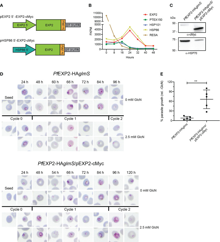
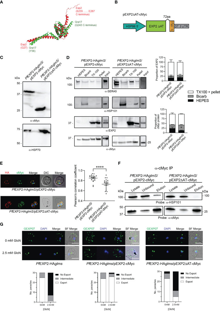
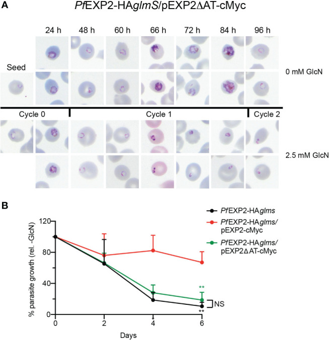
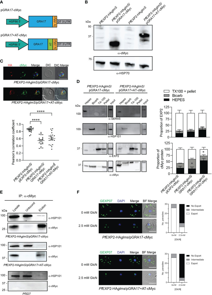
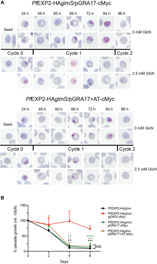
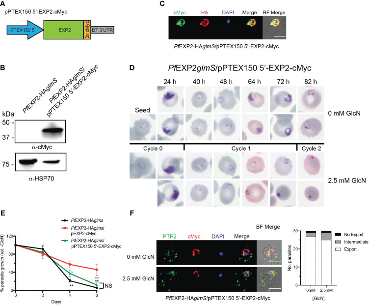
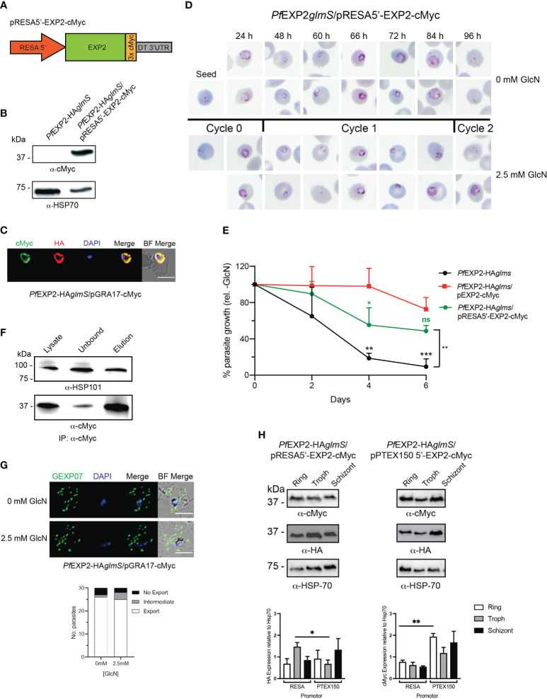
Apicomplexan parasites that reside within a parasitophorous vacuole harbor a conserved pore-forming protein that enables small-molecule transfer across the parasitophorous vacuole membrane (PVM). In Plasmodium parasites that cause malaria, this nutrient pore is formed by EXP2 which can complement the function of GRA17, an orthologous protein in Toxoplasma gondii. EXP2, however, has an additional function in Plasmodium parasites, serving also as the pore-forming component of the protein export machinery PTEX. To examine how EXP2 can play this additional role, transgenes that encoded truncations of EXP2, GRA17, hybrid GRA17-EXP2, or EXP2 under the transcriptional control of different promoters were expressed in EXP2 knockdown parasites to determine which could complement EXP2 function. This revealed that EXP2 is a unique pore-forming protein, and its protein export role in P. falciparum cannot be complemented by T. gondii GRA17. This was despite the addition of the EXP2 assembly strand and part of the linker helix to GRA17, which are regions necessary for the interaction of EXP2 with the other core PTEX components. This indicates that the body region of EXP2 plays a critical role in PTEX assembly and/or that the absence of other T. gondii GRA proteins in P. falciparum leads to its reduced efficiency of insertion into the PVM and complementation potential. Altering the timing and abundance of EXP2 expression did not affect protein export but affected parasite viability, indicating that the unique transcriptional profile of EXP2 when compared to other PTEX components enables it to serve an additional role in nutrient exchange.

Keywords: EXP2; GRA17; PTEX; Plasmodium; nutrient exchange; protein export.


Conflict of interest statement

The authors declare that the research was conducted in the absence of any commercial or financial relationships that could be construed as a potential conflict of interest. The author(s) declared that they were an editorial board member of Frontiers, at the time of submission. This had no impact on the peer review process and the final decision.

Figures

Figure 1
Figure 1
Complementation of PfEXP2-HAglmS with EXP2-cMyc. (A) Schematic of constructs to drive expression of a C-terminally cMyc-tagged EXP2 via the exp2 or hsp86 promoter. (B) Transcription analysis of the indicated genes across a single erythrocytic cycle. Data extracted from Otto et al. (Otto et al., 2010). FPKM, fragments per kilobase of exon per million mapped fragments. (C) Western blot analysis of parasite lysates confirms that PfEXP2-HAglmS/pEXP2-cMyc parasites express EXP2-cMyc (expected molecular weight is 39 kDa, with the 50-kDa band being non-specific labeling). HSP70 serves as a loading control. (D) Representative Giemsa-stained blood smears of three experiments where parasite lines were grown in the presence or absence of 2.5 mM GlcN after seeding at the stage indicated. (E) Growth of parasite lines after culturing for 10 days in the presence of 2.5 mM GlcN relative to untreated control as measured by Sybr Green 1 assay. Shown is the mean ± SD from five biological replicates, with significance determined by an unpaired t-test (**p < 0.01).
Figure 2
Figure 2
Complementation of PfEXP2-HAglmS with episomally expressed truncated EXP2ΔAT-cMyc. (A) Pymol alignment of the EXP2 structure (pdb 6E10 chain B residues 27–234) and the structured region of GRA17 (residues 55–243) generated using Alphafold. (B) Schematic of plasmid construct used for complementation. (C) Western blot analysis of parasite lysates confirms EXP2 is truncated in PfEXP2-HAglmS/EXP2ΔAT-cmyc (expected molecular weight is 30 kDa). (D) Representative Western blot from samples obtained from sequential solubility assays (left) and quantification of endogenous (upper graph) and cMyc-tagged protein (lower graph) in each fraction from three biological replicates. SERA5 is used as a marker for cytosolic proteins, HSP101 for membrane-associated proteins, and EXP2 for integral membrane proteins. Shown is the mean ± SEM from three biological replicates, with statistical significance determined by unpaired t-test (*p < 0.05). Note that input lanes were run at the same time but are shown as separate panels due to their location on the gel. (E) IFA of infected erythrocytes with DAPI staining (blue) marking the parasite’s nucleus (left), and the degree of co-localization between endogenous EXP2 (HA) and episomally expressed cMyc-tagged EXP2 calculated by measuring Pearson’s coefficients of merged Z-stack images of 20 cells (right). Shown is the mean ± SD, with significance determined by an unpaired t-test (****p < 0.001). (F) Western blots of whole-parasite lysate, unbound fraction and elution fraction following immunoprecipitation of cMyc-tagged proteins reveal that truncated EXP2 cannot interact with HSP101. (G) IFA of infected erythrocytes using antibodies to the Maurer’s cleft protein GEXP07 and quantification of GEXP07 localization (n = 30 cells from a single experiment), defined as either fully exported, not exported, or showing an intermediate export phenotype. Scale bar, 5 μm.
Figure 3
Figure 3
The acidic C-terminus of EXP2 is essential for parasite survival. (A) Representative Giemsa-stained blood smears of three experiments where PfEXP2-HAglmS/pEXP2ΔAT-cMyc parasites were grown in the presence or absence of 2.5 mM GlcN after seeding at the stage indicated. (B) Growth of the indicated parasite lines in the presence of 2.5 mM GlcN relative to untreated control as measured by Sybr Green 1 assay. Shown is the mean ± SD from three biological replicates. A one-way ANOVA with Dunnett’s test for multiple comparisons was used to determine whether differences in growth were significant, with that relative to PfEXP2-HAglmS/pEXP2-cMyc indicated (**p < 0.01); NS, not significant.
Figure 4
Figure 4
GRA17 with or without an acidic tail is unable to complement EXP2 function. (A) Schematic of GRA17 plasmid constructs used for complementation. (B) Western blot analysis of parasite lysates to confirm expression of GRA17-cMyc fusion proteins (expected molecular weight for GRA17-cMyc and GRA17-cMyc+AT is 31 and 40 kDa, respectively. (C) IFA of infected erythrocytes with DAPI staining (blue) marking the parasite’s nucleus (top), and the degree of co-localization between endogenous EXP2 (HA) and episomally expressed cMyc-tagged GRA17 calculated by measuring Pearson’s coefficients of merged Z-stack images of 20 cells from three experiments (bottom). Statistical significances were determined using one-way ANOVA with Welch correction and Dunnett’s test for multiple comparison (****p < 0.0001). (D) Representative Western blot of samples obtained from sequential solubility assays (left) and quantification of EXP2 (upper graph) and cMyc-tagged protein (lower graph) in each fraction from three biological replicates. SERA5 was used as a marker for cytosolic proteins, HSP101 for membrane-associated proteins, and EXP2 for integral membrane proteins. Shown is the mean ± SEM from three biological replicates, with statistical significance determined by unpaired t-test (*p < 0.05). Note that input lanes were run at the same time but are shown as separate panels due to their location on the gel (E) Western blots (n = 3) of whole-parasite lysate, unbound fraction and elution fraction following immunoprecipitation of cMyc-tagged proteins reveal that GRA17-cMyc proteins do not interact with HSP101. Lysates from Pf3D7 serve as a negative control. (F) IFA of infected erythrocytes using antibodies to the Maurer’s cleft protein GEXP07 and quantification of GEXP07 localization (n = 30 cells from a single experiment), defined as either fully exported, not exported, or showing an intermediate export phenotype. Scale bar, 5 μm.
Figure 5
Figure 5
GRA17 cannot complement the function of EXP2 even with addition of the EXP2 acidic tail. (A) Representative Giemsa-stained blood smears of three experiments where parasites were grown in the presence or absence of 2.5 mM GlcN after seeding at the stage indicated. (B) Growth of the indicated parasite lines in the presence of 2.5 mM GlcN relative to untreated control. Shown is the mean ± SD from three biological replicates. One-way ANOVA with Dunnett’s test for multiple comparisons was used to determine whether differences in growth were significant, with that relative to PfEXP2-HAglmS/pEXP2-cMyc indicated (**p < 0.01; ***p < 0.001; ****p < 0.0001); NS, not significant.
Figure 6
Figure 6
Expression of EXP2 under the ptex150 promoter affects parasite growth but not protein export. (A) Schematic of construct to drive expression of a C-terminally cMyc-tagged EXP2 via the ptex150 promoter. (B) Western blot analysis of parasite lysate to confirm expression of EXP2-cMyc (expected molecular weight is 39 kDa). (C) Representative IFA (n = 3) of infected erythrocytes with DAPI staining (blue) marking the parasite’s nucleus shows that endogenous EXP2 (HA) and cMyc-tagged EXP2 co-localize. (D) Representative Giemsa-stained blood smears of three experiments in which parasites were grown in the presence or absence of 2.5 mM GlcN. (E) Growth of the indicated parasite lines in the presence of 2.5 mM GlcN relative to untreated control. Shown is the mean ± SD from three biological replicates. One-way ANOVA with Dunnett’s test for multiple comparisons was used to determine whether differences in growth were significant, with that indicated relative to PfEXP2-HAglmS/pEXP2-cMyc (*p < 0.05; **p < 0.01); NS, not significant. (F) IFA of PfEXP2-HAglmS/pPTEX150 5′-EXP2-cMyc–infected erythrocytes using antibodies to the Maurer’s cleft protein PTP2 and quantification of PTP2 localization (n = 30 cells from a single experiment), defined as either fully exported, not exported, or showing an intermediate export phenotype. Scale bar, 5 μm.
Figure 7
Figure 7
Expression of EXP2 under the resa150 promoter. (A) Schematic of construct to drive expression of a C-terminally cMyc-tagged EXP2 via the resa promoter. (B) Western blot analysis of parasite lysate to confirm expression of EXP2-cMyc (expected molecular weight is 39 kDa). (C) Representative IFA (n = 3) of infected erythrocytes with DAPI staining (blue) marking the parasite’s nucleus (left) shows that endogenous EXP2 (HA) and cMyc-tagged EXP2 co-localize. (D) Representative Giemsa-stained blood smears of three experiments where parasites were grown in the presence or absence of 2.5 mM GlcN. (E) Growth of the indicated parasite lines in the presence of 2.5 mM GlcN relative to untreated control. Shown is the mean ± SD from three biological replicates. One-way ANOVA with Dunnett’s test for multiple comparisons was used to determine whether differences in growth were significant, with that relative to PfEXP2-HAglmS/pEXP2-cMyc indicated (*p < 0.05; **p < 0.01; ***p < 0.001; ns, not significant). (F) Western blots (n = 3) of whole-parasite lysate, unbound fraction and elution fraction following immunoprecipitation of cMyc-tagged protein shows that EXP2-cMyc interacts with HSP101. (G) IFA of infected erythrocytes using antibodies to the Maurer’s cleft protein GEXP07 and quantification of GEXP07 localization (n = 30 cells from a single experiment), defined as either fully exported, not exported, or showing an intermediate export phenotype. Scale bar, 5 μm. (H) Western blots of whole-parasite lysate from the indicated lines and quantitation of EXP2 (detected via anti-HA antibody) or episomally expressed EXP2 (detected anti-cMyc antibody) when expressed from either the resa or ptex150 promoter. Shown is the mean ± SEM from three biological replicates, with statistical significance determined by unpaired t-test (*p < 0.05; **p < 0.01).

Similar articles

Cited by

References

    1. Aikawa M., Torii M., Sjolander A., Berzins K., Perlmann P., Miller L. H. (1990). Pf155/RESA antigen is localized in dense granules of Plasmodium falciparum merozoites. Exp. Parasitol. 71, 326–329. doi: 10.1016/0014-4894(90)90037-D - DOI - PubMed
    1. Beck J. R., Muralidharan V., Oksman A., Goldberg D. E. (2014). HSP101/PTEX mediates export of diverse malaria effector proteins into the host erythrocyte. Nature 511, 592–595. doi: 10.1038/nature13574 - DOI - PMC - PubMed
    1. Brown G. V., Culvenor J. G., Crewther P. E., Bianco A. E., Coppel R. L., Saint R. B., et al. . (1985). Localization of the ring-infected erythrocyte surface antigen (RESA) of Plasmodium falciparum in merozoites and ring-infected erythrocytes. J. Exp. Med. 162, 774–779. doi: 10.1084/jem.162.2.774 - DOI - PMC - PubMed
    1. Bullen H. E., Charnaud S. C., Kalanon M., Riglar D. T., Dekiwadia C., Kangwanrangsan N., et al. . (2012). Biosynthesis, localisation and macromolecular arrangement of the Plasmodium falciparum translocon of exported proteins; PTEX. J. Biol. Chem. 287, 7871–7884. doi: 10.1074/jbc.M111.328591 - DOI - PMC - PubMed
    1. Charnaud S. C., Kumarasingha R., Bullen H. E., Crabb B. S., Gilson P. R. (2018). Knockdown of the translocon protein EXP2 in Plasmodium falciparum reduces growth and protein export. PloS One 13, e0204785. doi: 10.1371/journal.pone.0204785 - DOI - PMC - PubMed

Publication types

MeSH terms

Substances

LinkOut - more resources