. 2024 May;29(5):052918.
doi: 10.1117/1.JBO.29.5.052918. Epub 2024 Jan 27.

Speculum-free portable preterm imaging system

Affiliations

Speculum-free portable preterm imaging system

Tananant Boonya-Ananta et al. J Biomed Opt. 2024 May.

Abstract

Significance: Preterm birth is defined as a birth before 37 weeks of gestation and is one of the leading contributors to infant mortality rates globally. Premature birth can lead to life-long developmental impairment for the child. Unfortunately, there is a significant lack of tools to diagnose preterm birth risk, which limits patient care and the development of new therapies.

Aim: To develop a speculum-free, portable preterm imaging system (PPRIM) for cervical imaging; testing of the PPRIM system to resolve polarization properties of birefringent samples; and testing of the PPRIM under an IRB on healthy, non-pregnant volunteers for visualization and polarization analysis of cervical images.

Approach: The PPRIM can perform 4×3 Mueller-matrix imaging to characterize the remodeling of the uterine cervix during pregnancy. The PPRIM is built with a polarized imaging probe and a flexible insertable sheath made with a compatible flexible rubber-like material to maximize comfort and ease of use.

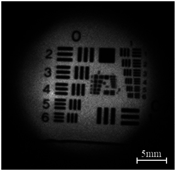
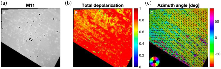
Results: The PPRIM device is developed to meet specific design specifications as a speculum-free, portable, and comfortable imaging system with polarized imaging capabilities. This system comprises a main imaging component and a flexible silicone inserter. The inserter is designed to maximize comfort and usability for the patient. The PPRIM shows high-resolution imaging capabilities at the 20 mm working distance and 25 mm circular field of view. The PPRIM demonstrates the ability to resolve birefringent sample orientation and full field capture of a healthy, non-pregnant cervix.

Conclusion: The development of the PPRIM aims to improve access to the standard of care for women's reproductive health using polarized Mueller-matrix imaging of the cervix and reduce infant and maternal mortality rates and better quality of life.

Keywords: Mueller-matrix; polarized imaging; portable device; pregnancy; preterm labor.


Figures

Fig. 1
Fig. 1
(a) Design assembly model render of PPRIM system; (b) exploded assembly of PPRIM indicating individual components; (c) exploded cross-section assembly of PPRIM, individual LED wire channels internal to the front housing, the lens optical train assembly and end housing internal features can be seen.
Fig. 2
Fig. 2
Explode assembly 2D drawing sectional view. Bill of materials can be seen in Table 1.
Fig. 3
Fig. 3
Inserter sheath dimension. All dimensions are in millimeters. The insertable length of the sheath is 65 mm, and the overall sheath length is 125 mm.
Fig. 4
Fig. 4
Overall dimensions of front housing with highlighted critical surfaces/features for design parameters. The red surface is critical for interface with the inserter sheath for a smooth insertion interface. The green surface is controlled for fitting into the back-end housing.
Fig. 5
Fig. 5
Front-end view of illumination and polarization components. (a) Model front view and (b) magnified single illumination pocket.
Fig. 6
Fig. 6
(a) Polarizer and LED pocket position at 45 deg increments, the blue rectangles indicate the position of two right circular polarizer/LED combination; (b) prototype model with fixed LEDs, polarizers, and quarter waveplates, and (c) model front view of illumination elements.
Fig. 7
Fig. 7
PPRIM prototype system with a flexible sheath. The main body casing is shown inserted into the flexible sheath.
Fig. 8
Fig. 8
3D SolidWorks model of casting mold for silicone sheath. The four main components of the casting mold include the two-mold casting body, the internal casting core, and the locking ring.
Fig. 9
Fig. 9
PPRIM inserter sheath silicone casting process images from casting mold model to 3D printed mold and the final silicone inserter product in blue.
Fig. 10
Fig. 10
Final optical design layout with light ray traces at 0.5in stock optics. (a) Zemax simulation results, (b) polychromatic diffraction simulation, and (c) field curvature simulation.
Fig. 11
Fig. 11
Image of a USAF target obtained with PPRIM.
Fig. 12
Fig. 12
(a) ZOE anatomical model with inserter, (b) the image of silicone cervix phantom removed from the model, and (c) raw PPRIM image of cervical phantom.
Fig. 13
Fig. 13
Birefringent silicone sample testing. The silicone sample is attached to a rotational stage under applied tension in a uniform direction. The stage is rotated to 80 deg at angular increments of 10 deg. (a) The azimuth angle of the sample indicates angular rotation from 10 deg (red) to 80 deg (cyan), as indicated by the color bar. (b) The plot indicates the sample’s mean angle and standard deviation at each rotational mount angle.
Fig. 14
Fig. 14
Excised birefringent Mueller-matrix decomposition. Chicken tendon sample aligned at 30  deg to the benchtop. (a) Intensity image M11, (b) total depolarization, and (c) azimuth angles. The black bars indicate the preferred alignment of the tissue within a 10-pixel range. Scale bar 5 mm.
Fig. 15
Fig. 15
In vivo human image samples from PPRIM polarized camera capture. Each column image is captured simultaneously and split into the corresponding polarization image channels. All images were taken with the right circular input state illumination. The first row (RH) is the horizontal polarization state, the second row (RV) is the vertical polarization state, the third row (RP) is the positive 45 deg, and the last row (RM) is the 45  deg polarization state. The scale bar in sample 4, RM image shows 5 mm.
Fig. 16
Fig. 16
In vivo human cervix sample showing (a) the intensity image, M11, (b) the retardation, and (c) the azimuth orientation, as shown by the azimuth angle. Scale bar 5 mm.

References

    1. Garfield R. E., et al. , “Methods and devices for the management of term and preterm labor,” Ann. N. Y. Acad. Sci. 943, 203–224 (2001).ANYAA910.1111/j.1749-6632.2001.tb03803.x - DOI - PubMed
    1. Soleimani F., Zaheri F., Abdi F., “Long-term neurodevelopmental outcomes after preterm birth,” Iran Red Crescent Med. J. 16(6), e17965 (2014).10.5812/ircmj.17965 - DOI - PMC - PubMed
    1. Saiga L. S., Doyle L., “An overview of mortality and sequelae of preterm birth from infancy to adulthood,” Lancet 371(9608), 261–269 (2008).LANCAO10.1016/S0140-6736(08)60136-1 - DOI - PubMed
    1. Grosse S. D., et al. , “Employer-sponsored plan expenditures for infants born preterm,” Pediatrics 140(4), e20171078 (2017).PEDIAU10.1542/peds.2017-1078 - DOI - PMC - PubMed
    1. Kozuki N., et al. , “The associations of birth intervals with small-for-gestational-age, preterm, and neonatal and infant mortality: a meta-analysis,” BMC Public Health 13 Suppl. 3(Suppl. 3), S3 (2013).10.1186/1471-2458-13-S3-S3 - DOI - PMC - PubMed

Publication types