. 2024 Jan 11:34:422-435.
doi: 10.1016/j.bioactmat.2023.12.027. eCollection 2024 Apr.

Targeted chemotherapy via HER2-based chimeric antigen receptor (CAR) engineered T-cell membrane coated polymeric nanoparticles

Affiliations

Targeted chemotherapy via HER2-based chimeric antigen receptor (CAR) engineered T-cell membrane coated polymeric nanoparticles

Serkan Yaman et al. Bioact Mater. .

Abstract

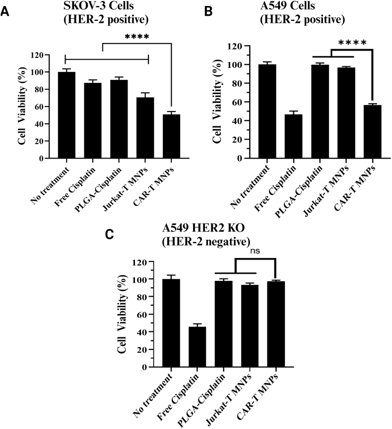
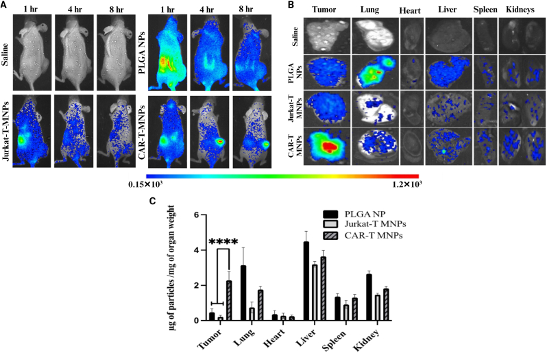
Cell membrane-derived nanoparticles (NPs) have recently gained popularity due to their desirable features in drug delivery such as mimicking properties of native cells, impeding systemic clearance, and altering foreign body responses. Besides NP technology, adoptive immunotherapy has emerged due to its promise in cancer specificity and therapeutic efficacy. In this research, we developed a biomimetic drug carrier based on chimeric antigen receptor (CAR) transduced T-cell membranes. For that purpose, anti-HER2 CAR-T cells were engineered via lentiviral transduction of anti-HER2 CAR coding lentiviral plasmids. Anti-HER2 CAR-T cells were characterized by their specific activities against the HER2 antigen and used for cell membrane extraction. Anti-cancer drug Cisplatin-loaded poly (D, l-lactide-co-glycolic acid) (PLGA) NPs were coated with anti-human epidermal growth factor receptor 2 (HER2)-specific CAR engineered T-cell membranes. Anti-HER2 CAR-T-cell membrane-coated PLGA NPs (CAR-T-MNPs) were characterized and confirmed via fluorescent microscopy and flow cytometry. Membrane-coated NPs showed a sustained drug release over the course of 21 days in physiological conditions. Cisplatin-loaded CAR-T-MNPs also inhibited the growth of multiple HER2+ cancer cells in vitro. In addition, in vitro uptake studies revealed that CAR-T-MNPs showed an increased uptake by A549 cells. These results were also confirmed via in vivo biodistribution and therapeutic studies using a subcutaneous lung cancer model in nude mice. CAR-T-MNPs localized preferentially at tumor areas compared to those of other studied groups and consisted of a significant reduction in tumor growth in tumor-bearing mice. In Conclusion, the new CAR modified cell membrane-coated NP drug-delivery platform has demonstrated its efficacy both in vitro and in vivo. Therefore, CAR engineered membrane-coated NP system could be a promising cell-mimicking drug carrier that could improve therapeutic outcomes of lung cancer treatments.

Keywords: CAR-T cells; Cancer chemotherapy; Membrane-based drug delivery; Nanoparticles.


Conflict of interest statement

There is no Conflict of Interest. The co-authors have approved the manuscript submission.

Figures

Image 1
Graphical abstract
Fig. 1
Fig. 1
Overview of proposed CAR-T-MNPs for targeted lung cancer therapy.
Fig. 2
Fig. 2
Confirmation of anti-HER2 scFv transduction on Jurkat T-cells by: A) Fluorescent microscopy imaging of mCherry expression on the transduced and sorted cells' brightfield channel (left) and fluorescent channel (right), (scalebar – 100 μm). B) Flow cytometric confirmation of Jurkat T-cell transduction, where anti-HER2 CAR transduced cells (red) show a distinctive shift against both mCherry and c-myc tag compared to the non-transduced Jurkat cell (black).
Fig. 3
Fig. 3
Physiochemical characterization of CAR-T-MNPs. A) TEM image of CAR-T-MNPs. B) Size, zeta potential, and polydispersity chart of NP formulations. C) Drug release kinetics of CAR-T-MNPs in PBS buffer. Anti-HER2 presence and functionality confirmation of: D) CAR-T-MNPs by flow cytometry against c-myc tag protein (CAR-T-MNPs (red) and uncoated PLGA NPs (Black)). E) CAR-T-cells by Western Blot shows a distinctive band correlating to anti-HER2 CAR scFv for CAR-T cells. No such band was observed for Jurkat-T-cells. (Number of replicates, n = 3).
Fig. 4
Fig. 4
Hemo- and cyto-compatibility of CAR-T-MNPs in vitro. A) Blood clotting effect of CAR-T-MNPs at different concentrations (250 μg/mL −1000 μg/mL). B) Hemolysis analysis of blood incubated with different concentrations (0 μg/mL - 1000 μg/mL) of CAR-T-MNPs. C) Cytocompatibility analysis of CAR-T-MNPs on AT1 cells to determine their in vitro cytotoxic effect on different concentrations (0 μg/mL −1000 μg/mL). (Number of replicates, n = 3).
Fig. 5
Fig. 5
Spectrophotometric analysis of different NP groups' uptake trend in: A) HER2-expressing A549 WT cells and B) A549 HER2 KO cells. C) Fluorescent uptake images of PLGA NPs, Jurkat-T-MNPs, and CAR-T-MNPs by A549 cells (500 μg/mL), NPs (green) and Nuclei (cyan). The data are shown as mean ± SD (Number of replicates, n = 4, ***P < 0.001, ****P < 0.0001).
Fig. 6
Fig. 6
In vitro therapeutic efficacy (cell killing) properties of CAR-T-MNPs. A) SKOV-3 B) A549 C) A549 HER2 KO cells exposed to free Cisplatin, PLGA NPs, Jurkat-T-MNPs & CAR-T-MNPs for 48 h. Cell viability was quantified via MTS assays after exposure (Number of replicates, n = 4, ****P < 0.0001).
Fig. 7
Fig. 7
In vivo biodistribution (targeting efficiency) studies. A)In vivo measurement of average color intensity of tumors/mm2 area on animal groups (CAR-T-MNPs, Jurkat-T-MNPs, PLGA NPs). B) Representative ex vivo organ images of all biodistribution study groups. C) Accumulation efficiency of fabricated nanoparticle systems in individual organs and tumors. (Number of replicates, n = 4, ****P < 0.0001).
Fig. 8
Fig. 8
In vivo therapeutic efficacy of CAR-T-MNPs on subcutaneous A549 tumor implanted in nude mice compared to Free Cisplatin, Jurkat-T-MNPs and saline. A) Tumor growth delay analysis for 2 weeks after treatment of mice with different groups. B) Animal survival analysis of tumor-implanted mice (sacrificed when tumor growth >20 mm in length/width). C) Histology analysis (H&E staining) of tumor tissue extracted from mice after experiment endpoint. (Number of replicates, n = 3–6, *P < 0.01).
Fig. 9
Fig. 9
In vivo toxicity analysis of CAR-T-MNPs on subcutaneous A549 tumor implanted in nude mice compared to Jurkat-T-MNPs and saline. A) Histological analysis of kidney tissue, immunohistochemistry of Ki67(green) and nucleus (blue). B) Quantitative analysis of Ki67 protein expression in kidney tissue. C) Masson’s trichrome staining of lung tissue comparing tissue scarring/collagen deposition as a result of cisplatin toxicity. (Number of replicates, n = 3, *P < 0.01).

Similar articles

Cited by

References

    1. Athey V.L., Suckling R.J., Tod A.M., Walters S.J., Rogers T.K. Early diagnosis of lung cancer: evaluation of a community-based social marketing intervention. Thorax. 2012;67(5):412–417. - PubMed
    1. Fukuoka M., Yano S., Giaccone G., Tamura T., Nakagawa K., Douillard J.Y., Nishiwaki Y., Vansteenkiste J., Kudoh S., Rischin D., Eek R., Horai T., Noda K., Takata I., Smit E., Averbuch S., Macleod A., Feyereislova A., Dong R.P., Baselga J. Multi-institutional randomized phase II trial of gefitinib for previously treated patients with advanced non-small-cell lung cancer (The IDEAL 1 Trial) [corrected] J. Clin. Oncol. 2003;21(12):2237–2246. - PubMed
    1. Siegel R., Naishadham D., Jemal A. Cancer statistics, 2013. CA A Cancer J. Clin. 2013;63(1):11–30. - PubMed
    1. Nurgali K., Jagoe R.T., Abalo R. Editorial: adverse effects of cancer chemotherapy: anything new to improve tolerance and reduce sequelae? Front. Pharmacol. 2018;9 - PMC - PubMed
    1. Dilalla V., Chaput G., Williams T., Sultanem K. Radiotherapy side effects: integrating a survivorship clinical lens to better serve patients. Curr. Oncol. 2020;27(2) - PMC - PubMed