Cerebral Arteriovenous Malformation With Ipsilateral Middle Cerebral Artery Occlusion: A Case Report
- PMID: 38283460
- PMCID: PMC10817826
- DOI: 10.7759/cureus.51193
Cerebral Arteriovenous Malformation With Ipsilateral Middle Cerebral Artery Occlusion: A Case Report
Abstract
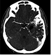
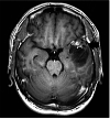
We report the case of a 29-year-old man who presented with a sudden headache. Computed tomography showed a small intraventricular hemorrhage in the left lateral ventricle. Cerebral angiograms suggested rupture of a coexisting feeder aneurysm in the left temporal cerebral arteriovenous malformation (AVM). The left proximal middle cerebral artery, a major feeding artery, was occluded near the AVM, with development of abnormal blood supply, such as in moyamoya-like vessels to the nidus. After endovascular embolization of the coexisting feeder aneurysm and feeding arteries, the patient underwent volume-staged Gamma Knife radiosurgery (GKS). Follow-up angiograms performed 4.5 years after the last GKS confirmed complete disappearance of the AVM. Around 4.8 years after GKS, the patient required surgical intervention to develop delayed cyst formation; however, the postoperative course was uneventful.
Keywords: cerebral arteriovenous malformation; cyst formation; feeder aneurysm; gamma knife radiosurgery; occlusion of the major feeding artery.
Copyright © 2023, Yada et al.
Conflict of interest statement
The authors have declared that no competing interests exist.
Figures


Similar articles
-
Gamma Knife surgical treatment for partially embolized cerebral arteriovenous malformations.J Neurosurg. 2016 Mar;124(3):767-76. doi: 10.3171/2015.1.JNS142711. Epub 2015 Aug 7. J Neurosurg. 2016. PMID: 26252461
-
Trigeminal Neuralgia from an Arteriovenous Malformation of the Trigeminal Root Entry Zone with a Flow-Related Feeding Artery Aneurysm: The Role of a Combined Endovascular and "Tailored" Surgical Treatment.Neurol India. 2021 May-Jun;69(3):744-747. doi: 10.4103/0028-3886.319235. Neurol India. 2021. PMID: 34169881
-
Acute rupture of a feeding artery aneurysm after embolization of a brain arteriovenous malformation.Interv Neuroradiol. 2015 Oct;21(5):613-9. doi: 10.1177/1591019915591740. Epub 2015 Jun 30. Interv Neuroradiol. 2015. PMID: 26126431 Free PMC article.
-
Ruptured de novo Aneurysm following Gamma Knife Surgery for Arteriovenous Malformation: Case Report.Stereotact Funct Neurosurg. 2017;95(6):379-384. doi: 10.1159/000481666. Epub 2017 Dec 1. Stereotact Funct Neurosurg. 2017. PMID: 29190619 Review.
-
Association of cerebral arteriovenous malformations and spontaneous occlusion of major feeding arteries: clinical and therapeutic implications.Neurosurgery. 1999 Nov;45(5):1105-11; discussion 1111-2. doi: 10.1097/00006123-199911000-00018. Neurosurgery. 1999. PMID: 10549926 Review.
References
-
- Defective cerebrovascular autoregulation in regions proximal to arteriovenous malformations of the brain: a case report and topic review. Solomon RA, Michelsen WJ. Neurosurgery. 1984;14:78–82. - PubMed
-
- Arteriovenous malformation in the territory of the occluded middle cerebral artery with massive intraoperative brain swelling: case report. Aoki N, Mizutani H. Neurosurgery. 1985;16:660–662. - PubMed
-
- Cerebral arteriovenous malformations associated with moyamoya phenomenon. Montanera W, Marotta TR, terBrugge KG, Lasjaunias P, Willinsky R, Wallace MC. https://www.ncbi.nlm.nih.gov/pmc/articles/PMC8332160/pdf/2124043.pdf. AJNR Am J Neuroradiol. 1990;11:1153–1156. - PMC - PubMed
-
- Association of cerebral arteriovenous malformations and spontaneous occlusion of major feeding arteries: clinical and therapeutic implications. Enam SA, Malik GM. Neurosurgery. 1999;45:1105–1111. - PubMed
Publication types
LinkOut - more resources
Full Text Sources
