

A Case of Spindle Cell Squamous Cell Carcinoma Manifesting in the Mandible Following Resection of Buccal Mucosal Squamous Cell Carcinoma
- PMID: 38283481
- PMCID: PMC10817819
- DOI: 10.7759/cureus.51191
A Case of Spindle Cell Squamous Cell Carcinoma Manifesting in the Mandible Following Resection of Buccal Mucosal Squamous Cell Carcinoma
Abstract
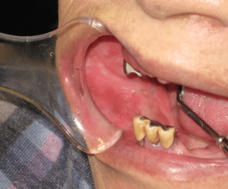
Spindle cell squamous cell carcinoma (SCSCC) represents a distinctive subtype of squamous cell carcinoma, characterized by a marked malignancy and sarcomatoid transformations predominantly comprising spindle-shaped cells. In this context, we executed a surgical resection of a buccal mucosal squamous cell carcinoma, encompassing the mandibular periosteum, for a case where buccal mucosal cancer had pervaded the mandibular gingival mucosa. Notably, in a period of one year and four months subsequent to this procedure, a spindle cell squamous cell carcinoma emerged as an intraosseous carcinoma, originating from the periosteum resection. This report delineates the occurrence of this rare pathology. The subject of this case is an 83-year-old female. She underwent a resection of a buccal mucosal squamous cell carcinoma, including the mandibular gingival periosteum, for cancer on the right buccal mucosa. The histopathological evaluation post-surgery confirmed the diagnosis of squamous cell carcinoma with clear margins. A computed tomography (CT) scan, conducted one year and four months postoperatively, disclosed a contrast-enhanced tumorous growth in the mandible. Owing to the considerable restriction in opening caused by scarring and the attendant challenges in biopsy acquisition, an expedited intraoperative diagnosis was rendered. This preliminary assessment indicated a spindle cell sarcoma, leading to a hemimandibular resection. The final histopathological diagnosis was spindle cell squamous cell carcinoma. Twelve months have elapsed since the surgical intervention, with no evidence of recurrence or metastasis observed to date.
Keywords: buccal mucosal squamous cell carcinoma; intraosseous carcinoma; mandible; spindle cell squamous cell carcinoma; transformation.
Copyright © 2023, Seta et al.
Conflict of interest statement
The authors have declared that no competing interests exist.
Figures


References
-
- A clinical study of oral spindle cell carcinoma after initial treatment of squamous cell carcinoma. Kobayashi Y, Fujii E, Iwaki H, et al. J Jpn Stomatol Soc. 2001;50:115–121.
-
- A case of tongue spindle cell carcinoma suspected of having transformed from squamous cell carcinoma after neoadjuvant chemotherapy [Japanese] Yamamoto T, Kondo E, Yamada S. Jpn J Oral Diag. 2017;30:289–294.
-
- A case of recurrent tongue cancer as spindle cell carcinoma following radiotherapy with intra-arterial chemotherapy [Japanese] Mitani S, Ukumori T, Tomidokoro Y, et al. Head Neck Cancer. 2014;40:334–337.
-
- A case of spindle cell carcinoma arising in the floor of the mouth after treatment of sqamous cell carcinoma on the contralateral side of the tongue. Miyajima Y, Hagiwara T, Furuya Y, et al. https://web.archive.org/web/20190506071348id_/https://www.jstage.jst.go.... Jpn J Oral Maxillofac Surg. 1998;45:187–189.
Publication types
LinkOut - more resources
Full Text Sources