. 2024 Jan 10:17:1294029.
doi: 10.3389/fncel.2023.1294029. eCollection 2023.

Impact of ferroptosis-related risk genes on macrophage M1/M2 polarization and prognosis in glioblastoma

Affiliations

Impact of ferroptosis-related risk genes on macrophage M1/M2 polarization and prognosis in glioblastoma

Xin Xu et al. Front Cell Neurosci. .

Abstract

Objective: To explore the effect impact of ferroptosis on macrophage polarization and patient prognosis in glioblastoma.

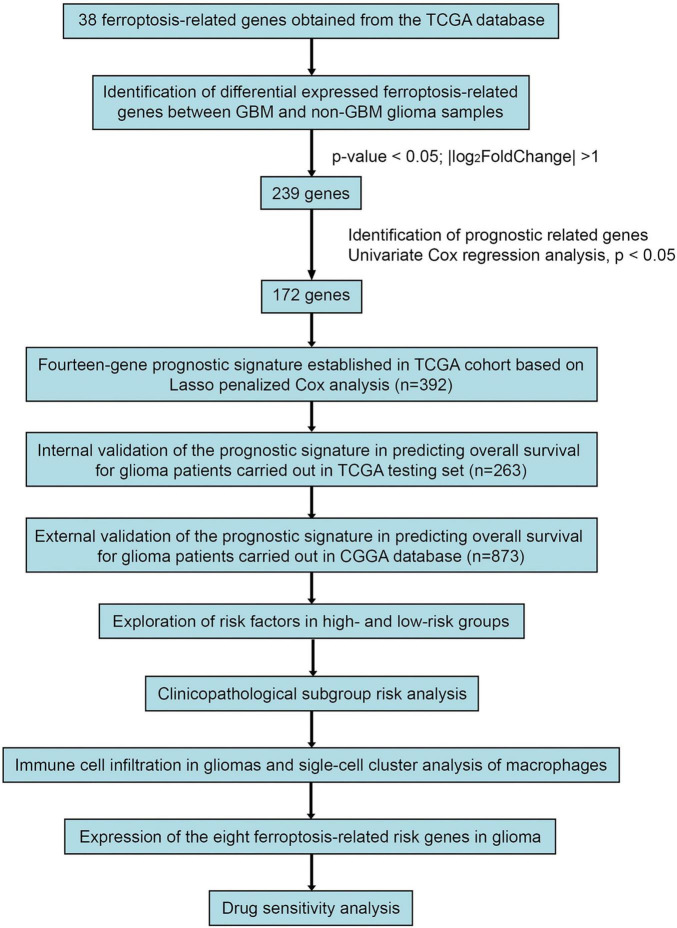
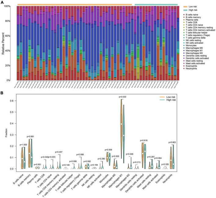
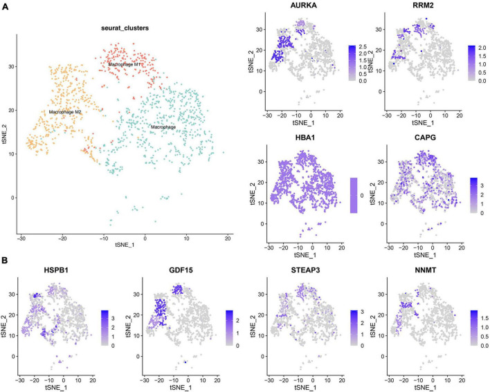
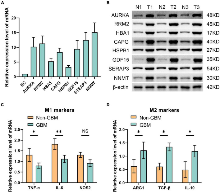
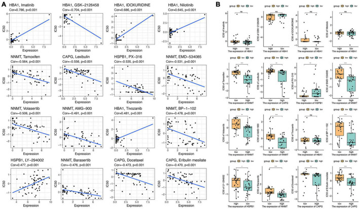
Methods: We screened ferroptosis-related risk from the public datasets of primary and recurrent glioblastoma, combined with reported ferroptosis genes, calculated the risk genes among the ferroptosis-related genes using the LASSO Cox regression model, and investigated the relationship between these ferroptosis-related risk genes in the tumor and the spectrum of infiltrating M1/M2 macrophages. Macrophages were analyzed using the CIBERSORTx deconvolution algorithm. Samples from The Cancer Genome Atlas (TCGA), Chinese Glioma Genome Atlas (CGGA) and a single-cell RNA sequencing dataset (GSE84465) were included. The expression levels of ferroptosis-related risk genes and molecular markers of M1 and M2 macrophages were detected by qPCR and western blot.

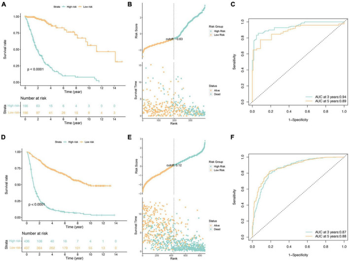
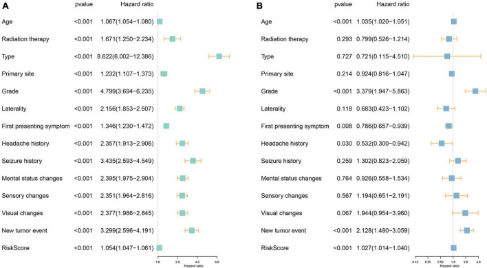
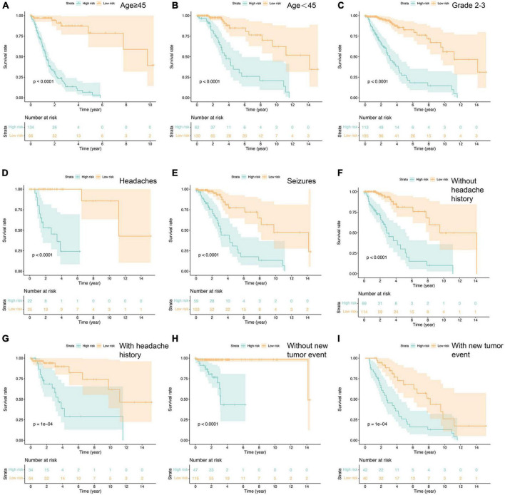
Results: A total of fourteen ferroptosis-related risk genes were obtained and the patients' risk scores were calculated. Compared with patients in the low-risk group, patients in the high-risk group had worse prognosis. The M1/M2 macrophage ratio and risk score were negatively correlated, indicating that the tumor microenvironment of glioblastoma in the high-risk group contained more M2 than M1 macrophages. In the single-cell RNA sequencing dataset, the risk score of ferroptosis-related genes in tumor cells was positively correlated with the proportion of high M2 macrophages. The expression of eight ferroptosis-related risk genes was increased in glioblastoma cell, which promoted the polarization of M1 macrophages to M2.

Conclusion: We investigated the fourteen ferroptosis-related risk genes in glioblastoma for the first time, and clarified the impact of ferroptosis-related risk genes on M1/M2 macrophage polarization and patient prognosis.

Keywords: M1/M2 macrophages; ferroptosis; glioblastoma; macrophage polarization; prognosis.


Conflict of interest statement

The authors declare that the research was conducted in the absence of any commercial or financial relationships that could be construed as a potential conflict of interest.

Figures

FIGURE 1
FIGURE 1
Study Workflow.
FIGURE 2
FIGURE 2
Survival analysis of glioma patients in the TCGA cohorts (A–C) and independent validation in the CGGA cohorts (D–F). (A,D) Kaplan-Meier survival analysis, (B,E) risk score by the fourteen ferroptosis-related risk genes, patients’ survival status and time, (C,F) time-dependent ROC curves. The 3 and 5-year AUCs were used to evaluate the prognostic accuracy, and the log-rank test was used to calculate the p-value.
FIGURE 3
FIGURE 3
Investigation of the independent prognostic risk factors in glioma patients. Univariate (A) and multivariate (B) Cox regression analysis of clinicopathologic characteristics were performed, and the hazard ratios (HR) and 95% confidence intervals (CI) were calculated, respectively.
FIGURE 4
FIGURE 4
Prognostic analysis in the subtypes with each independent risk factor group (A–I). P-value was calculated by log-rank test.
FIGURE 5
FIGURE 5
A total of 22 immune cell genotypes infiltration analysis in gliomas. (A) The proportions of immune cell subsets in the low- and high-risk groups were analyzed in the TCGA dataset. (B) The violin map showed statistical differences between the immune cells of low- and high-risk groups.
FIGURE 6
FIGURE 6
Single cell cluster analysis of macrophages in GBM. (A) The three identified macrophage classes in GBM. (B) The expression of eight ferroptosis-related risk genes in macrophage clusters.
FIGURE 7
FIGURE 7
Eight ferroptosis-related genes were detected by panel (A) qPCR at mRNA level and (B) Western blot at protein level. (C) qPCR was used to detect the molecular markers of M1 macrophages in non GBM and GBM groups. (D) The molecular markers of M2 macrophages in non GBM and GBM groups were detected by qPCR. *p < 0.05 and **p < 0.01.
FIGURE 8
FIGURE 8
Drug sensitivity analysis of eight ferroptosis-related genes. (A) The relationship between gene expression and the drug sensitivity for the 16 selected drugs. (B) The relationship between gene expression and drug sensitivities in low- and high-risk groups. NS means No significant, *p < 0.05, **p < 0.01, and ***p < 0.001.

Similar articles

Cited by

References

    1. Biswas S. K., Mantovani A. (2010). Macrophage plasticity and interaction with lymphocyte subsets: Cancer as a paradigm. Nat. Immunol. 11 889–896. 10.1038/ni.1937 - DOI - PubMed
    1. Chen C., Xie B., Li Z., Chen L., Chen Y., Zhou J., et al. (2022). Fascin enhances the vulnerability of breast cancer to erastin-induced ferroptosis. Cell Death Dis. 13:150. 10.1038/s41419-022-04579-1 - DOI - PMC - PubMed
    1. Chi H., Li B., Wang Q., Gao Z., Feng B., Xue H., et al. (2023). Opportunities and challenges related to ferroptosis in glioma and neuroblastoma. Front. Oncol. 13:1065994. 10.3389/fonc.2023.1065994 - DOI - PMC - PubMed
    1. Darmanis S., Sloan S. A., Croote D., Mignardi M., Chernikova S., Samghababi P., et al. (2017). Single-cell RNA-seq analysis of infiltrating neoplastic cells at the migrating front of human glioblastoma. Cell Rep. 21 1399–1410. - PMC - PubMed
    1. Fang Y., Zhang Z. (2020). Arsenic trioxide as a novel anti-glioma drug: A review. Cell Mol. Biol. Lett. 25:44. 10.1186/s11658-020-00236-7 - DOI - PMC - PubMed

LinkOut - more resources