. 2024 Jan 12:11:1310409.
doi: 10.3389/fcell.2023.1310409. eCollection 2023.

Microbiota affects mitochondria and immune cell infiltrations via alternative polyadenylation during postnatal heart development

Affiliations

Microbiota affects mitochondria and immune cell infiltrations via alternative polyadenylation during postnatal heart development

Xiang Liu et al. Front Cell Dev Biol. .

Abstract

There is a growing body of evidence supporting the significant impact of microbiota on heart development. Alternative polyadenylation (APA) is a crucial mechanism for gene expression regulation and has been implicated in postnatal heart development. Nonetheless, whether microbiota can influence postnatal heart development through the regulation of APA remains unclear. Therefore, we conducted APA sequencing on heart tissues collected from specific pathogen-free (SPF) mice and germ-free (GF) mice at three different developmental stages: within the first 24 h after birth (P1), 7-day-old SPF mice, and 7-day-old GF mice. This approach allowed us to obtain a comprehensive genome-wide profile of APA sites in the heart tissue samples. In this study, we made a significant observation that GF mice exhibited noticeably longer 3' untranslated region (3' UTR) lengths. Furthermore, we confirmed significant alterations in the 3' UTR lengths of mitochondria-related genes, namely Rala, Timm13, and Uqcc3. Interestingly, the GF condition resulted in a marked decrease in mitochondrial cristae density and a reduction in the level of Tomm20 in postnatal hearts. Moreover, we discovered a connection between Rala and Src, which further implicated their association with other differentially expressed genes (DEGs). Notably, most of the DEGs were significantly downregulated in GF mice, with the exceptions being Thbs1 and Egr1. Importantly, the GF condition demonstrated a correlation with a lower infiltration of immune cells, whereby the levels of resting NK cells, Th17 cells, immature dendritic cells, and plasma cells in GF mice were comparable to those observed in P1 mice. Furthermore, we established significant correlations between these immune cells and Rala as well as the related DEGs. Our findings clearly indicated that microbiota plays a vital role in postnatal heart development by affecting APA switching, mitochondria and immune cell infiltrations.

Keywords: alternative polyadenylation; heart development; immune cell; microbiota; mitochondrion.


Conflict of interest statement

The authors declare that the research was conducted in the absence of any commercial or financial relationships that could be construed as a potential conflict of interest.

Figures

FIGURE 1
FIGURE 1
The profile of APA switching in postnatal hearts with or without microbiota exposure. (A) The normalized 3′ UTR length of each gene in each sample was shown. Overall, the 3′ UTRs in P7 mice became longer compared to those in P1 mice, and P7_GF mice have even longer 3′ UTRs compared to the conventional counterparts. (B) Compared to P1 mice, there were 263 (178 longer and 85 shorter) genes harboring switched 3′ UTRs in P7 mice. (C) Compared to P1 mice, there were 238 (197 longer and 41 shorter) genes harboring switched 3′ UTRs in P7_GF mice. (D) Compared to P7 mice, there were 62 (44 longer and 18 shorter) genes harboring switched 3′ UTRs in P7_GF mice. (E) There were two overlapping genes, and 98, 97 and 26 genes were found exclusively in P1_vs_P7, P1_vs_P7_GF and P7_vs_P7_GF, respectively.
FIGURE 2
FIGURE 2
Functional annotations of the genes harboring switched 3′ UTRs. (A) Compared to P1 mice, genes with switched 3′ UTRs in P7 mice prominently enriched in the biological processes such as negative regulation of mRNA splicing via spliceosome, chromatin organization and actin filament-based process. (B) Compared to P1 mice, genes with switched 3′ UTRs in P7_GF mice were significantly related to protein catabolic process, actin filament-based movement and protein localization to organelle, etc. (C) Compared to P7 mice, genes with switched 3′ UTRs in P7_GF mice were notably enriched in myofibril assembly, mitochondrial membrane organization and Apelin signaling pathway, etc.
FIGURE 3
FIGURE 3
Verification of the APA switching genes related to mitochondria between P7 and P7_GF mice. (A) The functional annotations of genes with switched 3′ UTRs between P7 and P7_GF mice were visualized on Cytoscape. (B) The genes included in the module of mitochondrial membrane organization were selected for PPI analysis and Rala, Vamp2, Timm13 and Uqcc3 were identified as the key genes. (C) The 3′ UTR lengths of Rala, Vamp2, Timm13 and Uqcc3 were determined in sequencing. (D) The 3′ UTR lengths of Rala, Timm13 and Uqcc3 were further verified by qPCR. * p < 0.05, ** p < 0.01 versus the P1 mice; # p < 0.05, ## p < 0.01 versus the P7 mice. Data were expressed as mean ± SEM (n = 3).
FIGURE 4
FIGURE 4
Verification of the changes in myocardial mitochondria with or without microbiota exposure. (A) Postnatal hearts of GF mice showed an obvious reduction in mitochondrial cristae density. (B) Representative immunofluorescence images showing expressions of Tomm20 in postnatal hearts. DAPI indicates nuclei and Wheat Germ Agglutinin (WGA) highlights the cell membrane.
FIGURE 5
FIGURE 5
The profile of DEGs in postnatal hearts with or without microbiota exposure. (A) Compared to P1 mice, there were 988 (512 down and 476 up) DEGs in P7 mice. (B) Compared to P1 mice, there were 798 (421 down and 377 up) DEGs in P7_GF mice. (C) Compared to P7 mice, there were 79 (53 down and 26 up) DEGs in P7_GF mice. (D) There were two overlapping DEGs, and 379, 224 and 36 DEGs were found exclusively in P1_vs_P7, P1_vs_P7_GF and P7_vs_P7_GF, respectively. Upregulation and downregulation are presented as red and blue color in the Volcano plot, respectively.
FIGURE 6
FIGURE 6
Functional annotations and PPI analysis of the DEGs. (A, B) P1_vs_P7 and P1_vs_P7_GF shared many common functional GO terms, such as muscle tissue development, collagen-containing extracellular matrix and receptor ligand activity. (C) DEGs in P7_vs_P7_GF were significantly enriched in positive regulation of kinase activity, distal axon and transmembrane transporter binding, etc. (D) PPI analysis of the DEGs in P7_vs_P7_GF and the verified APA switching genes (Rala, Timm13 and Uqcc3) was conducted. It was found that only Rala can connect with another gene, namely Src, and then related to other DEGs. The top 5 GO terms are shown in the Bubble diagrams. The size of the dot represents gene count and the color illustrates adjusted p-value.
FIGURE 7
FIGURE 7
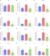
The expression levels of Rala and the related DEGs in sequencing. (A) The mRNA abundance of Rala remained unchanged between P7 and P7_GF mice. (B–J) Compared to P7 mice, the mRNA expressions of Src, Daam1, Fgr, Arrb2, Lrp5, Ube2m, Ighmbp2, Ptp4a3 and Bcl2l1 were notably downregulated. (K, L) Compared to P7 mice, the mRNA expressions of Thbs1 and Egr1 were dramatically upregulated. * p < 0.05, ** p < 0.01 versus the P1 mice; # p < 0.05, ## p < 0.01 versus the P7 mice. Data were expressed as mean ± SEM (n = 3).
FIGURE 8
FIGURE 8
The protein levels of Rala were differentially expressed by microbiota exposure. Representative immunofluorescence images showing expressions of Rala in postnatal hearts. DAPI indicates nuclei and Wheat Germ Agglutinin (WGA) highlights the cell membrane.
FIGURE 9
FIGURE 9
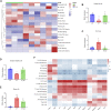
Immune cell infiltration and correlation analysis. (A) Overall, the three groups showed a difference in immune cells infiltrations, and the infiltrations in P7_GF mice appeared to be lower than those in the other two groups. Red represents high expression and blue low expression. (B) The resting NK cells were significantly decreased in P7 mice but completely restored in P7_GF mice. * p < 0.05 versus the P1 mice; # p < 0.05 versus the P7 mice. (C) The Th17 cells were notably increased in P7 mice but completely reversed in P7_GF mice. * p < 0.05 versus the P1 mice; # p < 0.05 versus the P7 mice. (D) The immature dendritic cells were significantly decreased in P7 mice. * p < 0.05 versus the P1 mice. (E) The plasma cells were significantly increased in P7 and P7_GF mice. * p < 0.05, ** p < 0.01 versus the P1 mice. (F) These immune cells were significantly correlated to Rala and the related DEGs. * p < 0.05, ** p < 0.01. Data were expressed as mean ± SEM (n = 3).

References

    1. Aurora A. B., Porrello E. R., Tan W., Mahmoud A. I., Hill J. A., Bassel-Duby R., et al. (2014). Macrophages are required for neonatal heart regeneration. J. Clin. Invest. 124, 1382–1392. 10.1172/JCI72181 - DOI - PMC - PubMed
    1. Barber D. F., Long E. O. (2003). Coexpression of CD58 or CD48 with intercellular adhesion molecule 1 on target cells enhances adhesion of resting NK cells. J. Immunol. 170, 294–299. 10.4049/jimmunol.170.1.294 - DOI - PubMed
    1. Chamberlain S. G., Owen D., Mott H. R. (2022). Membrane extraction by calmodulin underpins the disparate signalling of RalA and RalB. Bioessays 44, e2200011. 10.1002/bies.202200011 - DOI - PubMed
    1. Chen M., Lyu G., Han M., Nie H., Shen T., Chen W., et al. (2018). 3' UTR lengthening as a novel mechanism in regulating cellular senescence. Genome Res. 28, 285–294. 10.1101/gr.224451.117 - DOI - PMC - PubMed
    1. Chen Z., Huang A., Sun J., Jiang T., Qin F. X., Wu A. (2017). Inference of immune cell composition on the expression profiles of mouse tissue. Sci. Rep. 7, 40508. 10.1038/srep40508 - DOI - PMC - PubMed