. 2024 Jan 8;9(3):3866-3876.
doi: 10.1021/acsomega.3c07963. eCollection 2024 Jan 23.

Dye-Decolorizing Peroxidase of Streptomyces coelicolor (Sc DyPB) Exists as a Dynamic Mixture of Kinetically Different Oligomers

Affiliations

Dye-Decolorizing Peroxidase of Streptomyces coelicolor (Sc DyPB) Exists as a Dynamic Mixture of Kinetically Different Oligomers

Hegne Pupart et al. ACS Omega. .

Abstract

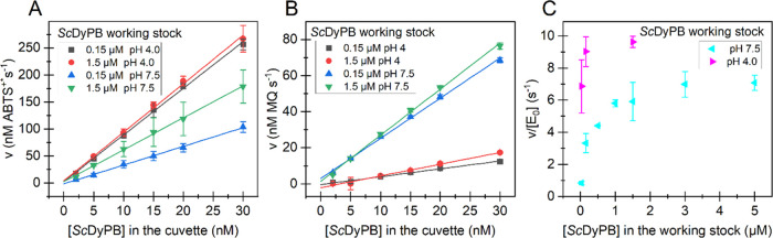
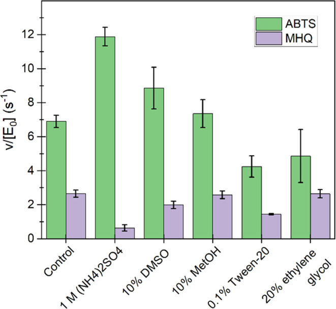
Dye-decolorizing peroxidases (DyPs) are heme-dependent enzymes that catalyze the oxidation of various substrates including environmental pollutants such as azo dyes and also lignin. DyPs often display complex non-Michaelis-Menten kinetics with substrate inhibition or positive cooperativity. Here, we performed in-depth kinetic characterization of the DyP of the bacterium Streptomyces coelicolor (ScDyPB). The activity of ScDyPB was found to be dependent on its concentration in the working stock used to initiate the reactions as well as on the pH of the working stock. Furthermore, the above-listed conditions had different effects on the oxidation of 2,2'-azino-di(3-ethyl-benzothiazoline-6-sulfonic acid) (ABTS) and methylhydroquinone, suggesting that different mechanisms are used in the oxidation of these substrates. The kinetics of the oxidation of ABTS were best described by the model whereby ScDyPB exists as a mixture of two kinetically different enzyme forms. Both forms obey the ping-pong kinetic mechanism, but one form is substrate-inhibited by the ABTS, whereas the other is not. Gel filtration chromatography and dynamic light scattering analyses revealed that ScDyPB exists as a complex mixture of molecules with different sizes. We propose that ScDyPB populations with low and high degrees of oligomerization have different kinetic properties. Such enzyme oligomerization-dependent modulation of the kinetic properties adds further dimension to the complexity of the kinetics of DyPs but also suggests novel possibilities for the regulation of their catalytic activity.


Conflict of interest statement

The authors declare no competing financial interest.

Figures

Scheme 1
Scheme 1. Oxidation of 2,2′-Azino-di-(3-ethyl-benzothiazoline-6-sulfonic acid) (ABTS) and methylhydroquinone (MHQ)
One-electron oxidation of ABTS to the ABTS cation radical and two-electron oxidation of MHQ to methylquinone (MQ) (absorption maxima at 420 and 251 nm, respectively).
Figure 1
Figure 1
Dependency of initial rates of reducing substrate oxidation on pH by ScDyPB. The time curves of the oxidation of (A) ABTS and (B) MHQ. The pH values are given in the plot. (C) Dependency of the initial rates (measured between 30 and 60 s) of the oxidation of ABTS and MHQ on pH. All reactions were performed at 25 °C. Buffers were 50 mM sodium citrate (for pH 3.0 and 3.5), 50 mM sodium acetate (for pH 4.0–5.0), and 50 mM Bis–Tris–HCl (for pH 5.5 and 6.0). Reactions were initiated by the addition of the ScDyPB from its working stock (1.5 μM ScDyPB in 20 mM Tris-HCl pH 7.5 supplemented with 0.1 g L–1 BSA and 0.1 M NaCl) to the cuvette containing the mixture of the substrate and H2O2. The final concentration of the ScDyPB in the cuvette was 15 nM. The oxidation of ABTS was measured using 1 mM ABTS and 100 μM H2O2. The oxidation of MHQ was measured using 1 mM MHQ (note that MHQ preparation had a high background absorbance at pH 6.0) and 1 mM H2O2. Data are presented as average values (n = 3, independent experiments), and error bars show SD. For clarity, the error bars are not shown for the traces in panels (A) and (B).
Figure 2
Figure 2
Dependency of the activity on the concentration of ScDyPB in the cuvette and in its working stock. All reactions were performed in NaAc buffer (50 mM, pH 4.0) at 25 °C. The oxidation of ABTS was measured using 1 mM ABTS and 100 μM H2O2 and the oxidation of MHQ with 1 mM MHQ and 1 mM H2O2. The rates of oxidation of (A) ABTS and (B) MHQ at different concentrations of ScDyPB in the cuvette. The concentration of ScDyPB in its working stock and the pH of the working stock are shown in the figure. Solid lines show the linear regression of the data. (C) Dependency of the ABTS oxidizing activity of ScDyPB on its concentration in the working stock. The pH of the ScDyPB working stock is shown in the figure. The concentration of the ScDyPB in the cuvette was 15 nM. The working stocks of ScDyPB, with concentrations of 30 nM–5 μM, were prepared in 20 mM Tris pH 7.5, 0.1 M NaCl, or 50 mM NaAc pH 4.0 buffers (both supplemented with 0.1 g L–1 BSA). Data are presented as average values (n = 3, independent experiments), and error bars show SD.
Figure 3
Figure 3
Effects of different additives in the working stock of ScDyPB on the ABTS and MHQ oxidizing activity. Prior to the activity measurements, the ScDyPB working stock (1.5 μM ScDyPB in 20 mM Tris-HCl pH 7.5 supplemented with 0.1 g L–1 BSA and 0.1 M NaCl) was incubated for 30 min at 25 °C in the presence of different compounds as indicated in the figure. The activity was measured in 50 mM NaAc at pH 4.0 using 15 nM ScDyPB (100-fold dilution of the working stock to the cuvette). The activity was measured using 1 mM ABTS and 100 μM H2O2 or 1 mM MHQ and 1 mM H2O2. Data are presented as average values (n = 3, independent experiments), and error bars show SD.
Figure 4
Figure 4
Kinetics of the oxidation of ABTS by ScDyPB. Dependency of initial rates of the oxidation of ABTS on the concentration of (A) ABTS and (B) H2O2 and the dependency of apparent parameters (C) kcatapp, (D) KM(H2O2)app, and (E) kcatapp/KM(H2O2)app on the concentration of ABTS. Reactions were made in 50 mM NaAc (pH 4.0) at 25 °C. The concentration of ScDyPB was 15 nM, and the reactions were initiated by the addition of ScDyPB from the working stock with 1.5 μM ScDyPB in 20 mM Tris pH 7.5 (supplemented with 0.1 g L–1 BSA and 0.1 M NaCl). Solid lines in (A) and (B) show the global nonlinear regression of the data according to eq 2. The concentration of the substrate that has been kept constant within the series is indicated in the plot. The values of apparent kinetic parameters for H2O2 shown in panels (C–E) were derived by nonlinear regression analysis of the data in panel (B) according to the Michaelis–Menten equation (eq S1, for the fit, see Figure S2B). Data are presented as average values (n = 3, independent experiments), and error bars show SD.
Figure 5
Figure 5
Analysis of the size distribution of ScDyPB. (A) Gel filtration chromatogram of ScDyPB in 20 mM Tris-HCl (pH, 7.5) (supplemented with 100 mM NaCl). Black squares show the elution volume of standard proteins: ferritin (440 kDa), aldolase (158 kDa), conalbumin (75 kDa), and ovalbumin (44 kDa). The red line shows linear regression analysis of the mobility of the standard proteins used for calibration. The red square shows the expected elution volume of the ScDyPB monomer. (B, C) Dynamic light scattering (DLS) analysis of 1.5 μM ScDyPB in 50 mM NaAc pH 4.0, in 20 mM Tris pH 7.5, and in 20 mM Tris pH 7.5 supplemented with 1.0 M ammonium sulfate (as indicated in the plot). Size distribution based on the intensity (B) or volume (C). Traces show the average of at least four consecutive scans.
Figure 6
Figure 6
Structure of ScDyPB and simplest possible mechanism of the formation of kinetically different forms of the enzyme. (A) Structure of ScDyPB (PDB: 4GU7). The structure of the ScDyPB monomer, where α- helices, β-sheets, and loops are colored blue, pink, and tan, respectively. The heme is shown as a stick model. Nitrogen, oxygen, and iron atoms are colored blue, red, and brown, respectively. The inset shows the heme access channel of ScDyPB, the propionate pocket (left). The ScDyPB hexamer (trimer of dimers), with dimers shown in different colors and the approximate dimensions of the hexamer (right). (B) Simplest mechanism of the catalysis by ScDyPB that explains the experimental observations of this study. ScDyPB exists as an equilibrium (slow compared with the time frame used for the activity measurements) of the enzyme forms with low (LDO form) and high degrees of oligomerization (HDO form). For the simplicity of visualization of the concept, the LDO- and HDO forms are represented by the monomer and dimer, respectively (in a real system, the degree of oligomerization of both forms is much higher). In their resting state (RS), both forms of the enzyme can be oxidized by H2O2 to form an active intermediate, compound I (Cpd I). In the case of ABTS as the reducing substrate, the RS is restored by the long-range electron transfer to Cdp I. For MHQ, there is no surface binding site, and Cpd I is reduced via direct electron transfer to the heme iron. The heme access channel is assumed to be inaccessible for the reducing substrate in the enzyme in its HDO form. Therefore, the oxidation of MHQ occurs only with the enzyme in its LDO form. Substrate inhibition by ABTS results from the nonproductive binding of ABTS to the heme (NP) and is possible only with the enzyme in its LDO form. Thus, the conditions that favor the HDO form of ScDyPB will decrease the MHQ oxidizing activity through blockage of the access to the heme and increase ABTS oxidizing through relieving substrate inhibition. Note that possible substrate inhibition by MHQ and oxidation of ABTS through direct electron transfer do not change the general outcome of the model and are omitted for simplicity.

References

    1. Kim S. J.; Shoda M. Purification and characterization of a novel peroxidase from Geotrichum candidum Dec 1 involved in decolorization of dyes. Appl. Environ. Microbiol. 1999, 65, 1029–1035. 10.1128/AEM.65.3.1029-1035.1999. - DOI - PMC - PubMed
    1. Scheibner M.; Hülsdau B.; Zelena K.; Nimtz M.; de Boer L.; Berger R. G.; Zorn H. Novel peroxidases of Marasmius scorodonius degrade β-carotene. Appl. Microbiol. Biotechnol. 2008, 77, 1241–1250. 10.1007/s00253-007-1261-9. - DOI - PubMed
    1. van Bloois E.; Pazmiño D. E. T.; Winter R. T.; Fraaije M. W. A robust and extracellular heme-containing peroxidase from Thermobifida fusca as prototype of a bacterial peroxidase superfamily. Appl. Microbiol. Biotechnol. 2010, 86, 1419–1430. 10.1007/s00253-009-2369-x. - DOI - PMC - PubMed
    1. Singh R.; Eltis L. D. The multihued palette of dye-decolorizing peroxidases. Arch. Biochem. Biophys. 2015, 574, 56–65. 10.1016/j.abb.2015.01.014. - DOI - PubMed
    1. Yoshida T.; Sugano Y. Unexpected diversity of dye-decolorizing peroxidases. Biochem. Biophys. Rep. 2023, 33, 10140110.1016/j.bbrep.2022.101401. - DOI - PMC - PubMed

LinkOut - more resources