. 2024 Jan 9;9(3):4096-4101.
doi: 10.1021/acsomega.3c09288. eCollection 2024 Jan 23.

Abundance of G-Quadruplex Forming Sequences in the Hepatitis Delta Virus Genomes

Affiliations

Abundance of G-Quadruplex Forming Sequences in the Hepatitis Delta Virus Genomes

Václav Brázda et al. ACS Omega. .

Abstract

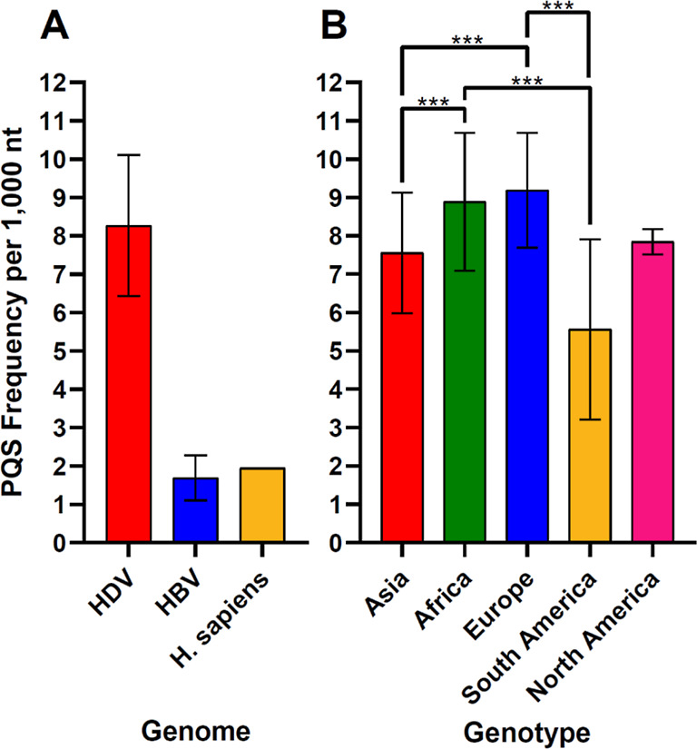
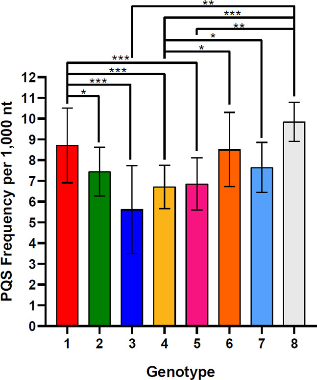
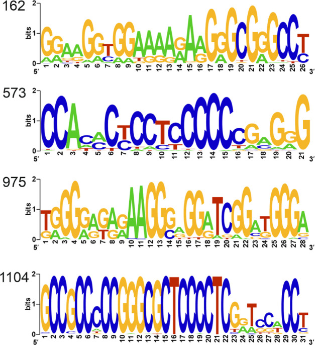
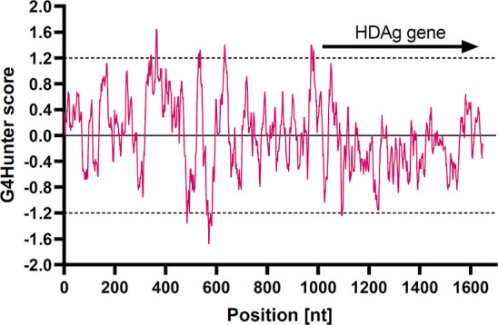
Hepatitis delta virus (HDV) is a highly unusual RNA satellite virus that depends on the presence of hepatitis B virus (HBV) to be infectious. Its compact and variable single-stranded RNA genome consists of eight major genotypes distributed unevenly across different continents. The significance of noncanonical secondary structures such as G-quadruplexes (G4s) is increasingly recognized at the DNA and RNA levels, particularly for transcription, replication, and translation. G4s are formed from guanine-rich sequences and have been identified in the vast majority of viral, eukaryotic, and prokaryotic genomes. In this study, we analyzed the G4 propensity of HDV genomes by using G4Hunter. Unlike HBV, which has a G4 density similar to that of the human genome, HDV displays a significantly higher number of potential quadruplex-forming sequences (PQS), with a density more than four times greater than that of the human genome. This finding suggests a critical role for G4s in HDV, especially given that the PQS regions are conserved across HDV genotypes. Furthermore, the prevalence of G4-forming sequences may represent a promising target for therapeutic interventions to control HDV replication.


Conflict of interest statement

The authors declare no competing financial interest.

Figures

Figure 1
Figure 1
Comparison of PQS frequencies (A) in the HDV, HBV, and human genomes; (B) in HDV genomes isolated in different continents. Significant differences are shown by asterisks: *p-value < 0.05; **p-value < 0.01; ***p-value < 0.001.
Figure 2
Figure 2
Comparison of the PQS frequencies in HDV genotypes. Significant differences are shown by asterisks: *p-value < 0.05; **p-value < 0.01; ***p-value < 0.001.
Figure 3
Figure 3
Conservation of bases in the selected G4s. LOGO representation of the consensus PQS motifs in four locations (indicated on the left) with the highest G4Hunter scores.
Figure 4
Figure 4
PQSs location within the reference HDV genome (NC_076103.1). The regions identified as PQS by G4Hunter, have either positive scores exceeding 1.2 (above the dashed line) or negative scores (below −1.2, PQS in the complementary antigenome sequence). The coding region is shown by arrow.

Similar articles

Cited by

References

    1. Koff R. S. Fulminant Hepatitis Due to HBV/HDV Coinfection. Hosp. Pract. 1987, 22 (11), 123–154. 10.1080/21548331.1987.11703365. - DOI - PubMed
    1. Abbas Z.; Qureshi M.; Hamid S.; Jafri W. Hepatocellular Carcinoma in Hepatitis D: Does It Differ from Hepatitis B Monoinfection?. Saudi J. Gastroenterol. 2012, 18 (1), 18–22. 10.4103/1319-3767.91731. - DOI - PMC - PubMed
    1. Alves C.; Branco C.; Cunha C. Hepatitis Delta Virus: A Peculiar Virus. Adv. Virol. 2013, 2013, 1–11. 10.1155/2013/560105. - DOI - PMC - PubMed
    1. Zi J.; Gao X.; Du J.; Xu H.; Niu J.; Chi X. Multiple Regions Drive Hepatitis Delta Virus Proliferation and Are Therapeutic Targets. Front. Microbiol. 2022, 13, 838382.10.3389/fmicb.2022.838382. - DOI - PMC - PubMed
    1. Tseng C.-H.; Lai M. M. C. Hepatitis Delta Virus RNA Replication. Viruses 2009, 1 (3), 818–831. 10.3390/v1030818. - DOI - PMC - PubMed

LinkOut - more resources