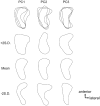
Morphometric Analysis of the Tibial Attachment Shape of the Anterior Cruciate Ligament and Its Relationship With the Location of the Anterior Horn of the Lateral Meniscus
- PMID: 38284162
- PMCID: PMC10905983
- DOI: 10.1177/03635465231219978
Morphometric Analysis of the Tibial Attachment Shape of the Anterior Cruciate Ligament and Its Relationship With the Location of the Anterior Horn of the Lateral Meniscus
Abstract
Background: The success of anterior cruciate ligament (ACL) reconstruction relies on the accurate replication of the native ACL anatomy, including attachment shapes. The tibial attachment of the ACL exhibits significant shape variations with elliptical, C, and triangular shapes, highlighting the need for objective classification methods and additional information to identify individual anatomic variations.
Hypothesis: The location of the attachment of the anterior horn of the lateral meniscus (AHLM) may determine the shape of the ACL attachment.
Study design: Cross-sectional study; Level of evidence, 3.
Methods: The study used 25 knees from 17 Japanese cadavers for macroscopic anatomic examination and quantitative analysis. The shape of the ACL attachment was quantified using principal component analysis with elliptical Fourier descriptors, whereas the AHLM location was quantified by measuring its mediolateral and anteroposterior positions on the superior surface of the tibia. Reliability was assessed statistically.
Results: The shape of the tibial attachment of the ACL varied among individuals and was classified as elliptical, C-shaped, or triangular. Scatterplots of the principal components of the ACL attachment shape showed overlapping regions of elliptical, C-shaped, and triangular ACL attachments, indicating that a C-shaped attachment is intermediate between elliptical and triangular attachments. The location of the AHLM attachment also varied, with areas in the anterolateral, anteromedial, or posteromedial region. The ACL shape and AHLM location were related, with elliptical, C-shaped, and triangular ACL attachments corresponding to anterolateral, anteromedial, and posteromedial AHLM attachments, respectively.
Conclusion: The AHLM attachment location influences the shape of the ACL attachment. Information on the location of the AHLM attachment can aid in predicting the shape of the ACL attachment during ACL reconstruction, potentially improving footprint coverage.
Keywords: anatomy; anterior cruciate ligament; lateral meniscus.
Conflict of interest statement
The authors declared that they have no conflicts of interest in the authorship and publication of this contribution. AOSSM checks author disclosures against the Open Payments Database (OPD). AOSSM has not conducted an independent investigation on the OPD and disclaims any liability or responsibility relating thereto.
Figures


Similar articles
-
Anterior cruciate ligament tibial insertion site is elliptical or triangular shaped in healthy young adults: high-resolution 3-T MRI analysis.Knee Surg Sports Traumatol Arthrosc. 2018 Feb;26(2):485-490. doi: 10.1007/s00167-017-4607-6. Epub 2017 Jun 24. Knee Surg Sports Traumatol Arthrosc. 2018. PMID: 28647841
-
Tibial insertions of the anterior cruciate ligament and the anterior horn of the lateral meniscus: A histological and computed tomographic study.Knee. 2017 Aug;24(4):782-791. doi: 10.1016/j.knee.2017.04.014. Epub 2017 May 27. Knee. 2017. PMID: 28559005
-
Tibial insertion of the anterior cruciate ligament and anterior horn of the lateral meniscus share the lateral slope of the medial intercondylar ridge: A computed tomography study in a young, healthy population.Knee. 2019 Jun;26(3):612-618. doi: 10.1016/j.knee.2019.04.009. Epub 2019 May 9. Knee. 2019. PMID: 31078391
-
ACL anatomy: Is there still something to learn?Rev Esp Cir Ortop Traumatol. 2024 Jul-Aug;68(4):T422-T427. doi: 10.1016/j.recot.2024.03.009. Epub 2024 Mar 18. Rev Esp Cir Ortop Traumatol. 2024. PMID: 38508380 Review. English, Spanish.
-
ACL anatomy: Is there still something to learn?Rev Esp Cir Ortop Traumatol. 2024 Jul-Aug;68(4):422-427. doi: 10.1016/j.recot.2023.02.005. Epub 2023 Feb 12. Rev Esp Cir Ortop Traumatol. 2024. PMID: 36787832 Review. English, Spanish.
References
-
- Colombet P, Robinson J, Christel P, et al.. Morphology of anterior cruciate ligament attachments for anatomic reconstruction: a cadaveric dissection and radiographic study. Arthroscopy. 2006;22(9):984-992. - PubMed
-
- de Abreu-e-Silva GM, de Oliveira MH, Maranhao GS, et al.. Three-dimensional computed tomography evaluation of anterior cruciate ligament footprint for anatomic single-bundle reconstruction. Knee Surg Sports Traumatol Arthrosc. 2015;23(3):770-776. - PubMed
-
- Ferretti M, Doca D, Ingham SM, Cohen M, Fu FH. Bony and soft tissue landmarks of the ACL tibial insertion site: an anatomical study. Knee Surg Sports Traumatol Arthrosc. 2012;20(1):62-68. - PubMed
-
- Fujishiro H, Tsukada S, Nakamura T, et al.. Attachment area of fibres from the horns of lateral meniscus: anatomic study with special reference to the positional relationship of anterior cruciate ligament. Knee Surg Sports Traumatol Arthrosc. 2017;25(2):368-373. - PubMed
MeSH terms
LinkOut - more resources
Full Text Sources

