. 2024 Jan;102(1):e25278.
doi: 10.1002/jnr.25278.

A phenotypically robust model of spinal and bulbar muscular atrophy in Drosophila

Affiliations

A phenotypically robust model of spinal and bulbar muscular atrophy in Drosophila

Kristin Richardson et al. J Neurosci Res. 2024 Jan.

Abstract

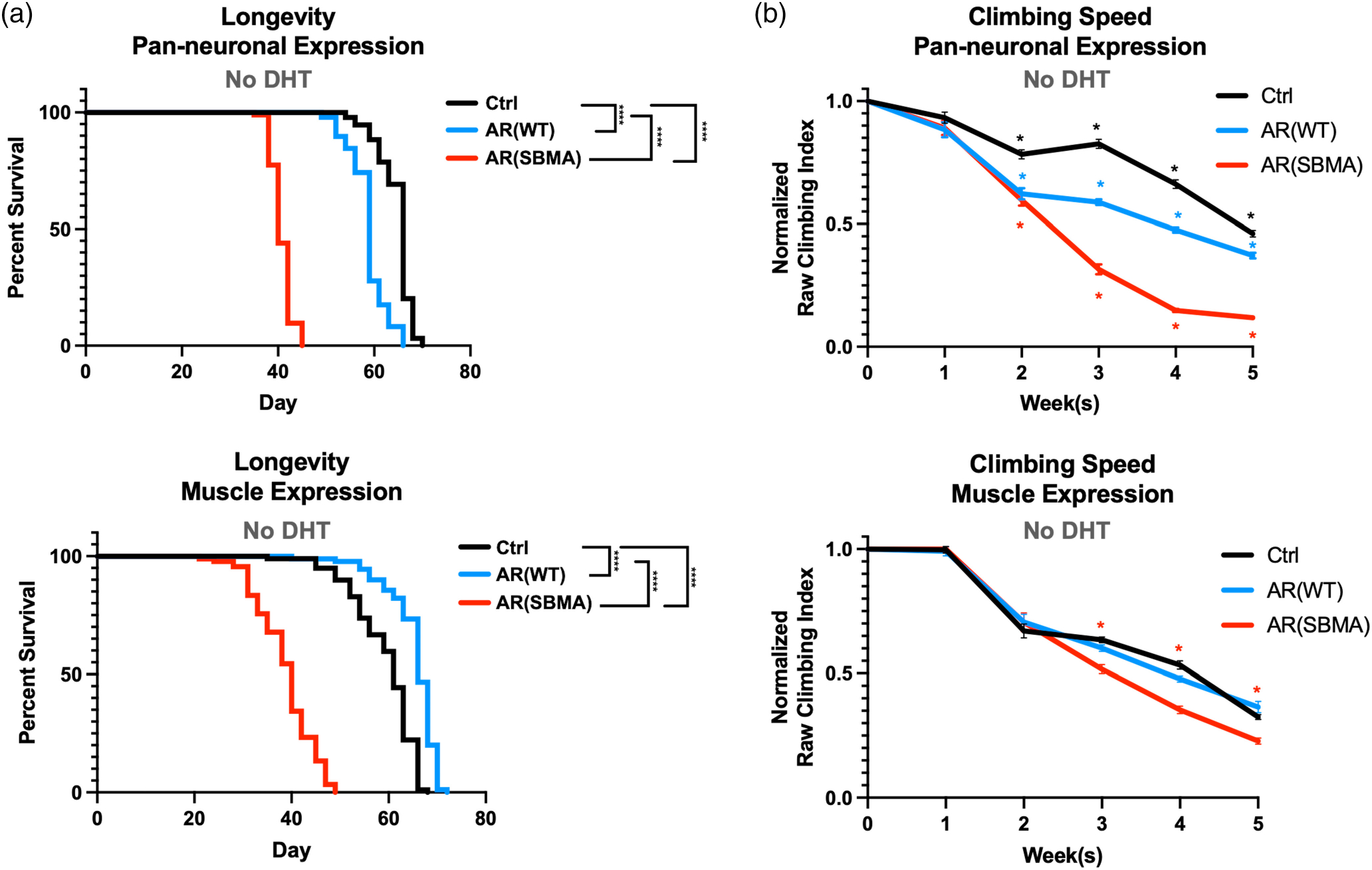
Spinal and bulbar muscular atrophy (SBMA) is an X-linked disorder that affects males who inherit the androgen receptor (AR) gene with an abnormal CAG triplet repeat expansion. The resulting protein contains an elongated polyglutamine (polyQ) tract and causes motor neuron degeneration in an androgen-dependent manner. The precise molecular sequelae of SBMA are unclear. To assist with its investigation and the identification of therapeutic options, we report here a new model of SBMA in Drosophila melanogaster. We generated transgenic flies that express the full-length, human AR with a wild-type or pathogenic polyQ repeat. Each transgene is inserted into the same safe harbor site on the third chromosome of the fly as a single copy and in the same orientation. Expression of pathogenic AR, but not of its wild-type variant, in neurons or muscles leads to consistent, progressive defects in longevity and motility that are concomitant with polyQ-expanded AR protein aggregation and reduced complexity in neuromuscular junctions. Additional assays show adult fly eye abnormalities associated with the pathogenic AR species. The detrimental effects of pathogenic AR are accentuated by feeding flies the androgen, dihydrotestosterone. This new, robust SBMA model can be a valuable tool toward future investigations of this incurable disease.

Keywords: genetics; muscle; neuron; polyglutamine; triplet repeat.


Conflict of interest statement

CONFLICT OF INTEREST STATEMENT

The authors declare that the research was conducted in the absence of any commercial or financial relationships that could be construed as a potential conflict of interest.

Figures

FIGURE 1
FIGURE 1
New transgenic fly lines for SBMA. (a) Amino acid sequence of the human AR protein used. The polyQ is encoded by a CAG/CAA doublet repeat to circumvent the possibility of mRNA- and non-AUG translation-dependent toxicity. Bolded: HA epitope tag. (b) Insertion strategy into site VK00033 on chromosome 3 of the fly. (c) Western blotting from adult flies expressing the noted transgenes. Whole fly lysates. elav-Gal4: pan-neuronal expression. Mef2- Gal4: muscle cell expression. Black arrows: main AR bands; solid red bracketed line: SDS-resistant, polyQ-expanded AR trapped in the stacking gel; dotted, red bracketed line, polyQ-expanded AR trapped in resolving gel; gray bracketed line: likely proteolytic products of AR. Each lane is from an independent repeat.
FIGURE 2
FIGURE 2
Longevity and motility outcomes from expression of W T or polyQ-expanded AR in neuronal or muscle cells. (a) Longevity assays of female flies expressing the noted transgenes. No DHT was introduced at any point of their lives. N ≧ 100 flies per group. ****p < .0 0 01, log-rank tests. (b) Normalized motility assays of female flies expressing the noted transgenes in neurons or muscles. N ≧ 100 flies per group *p < .05. Statistical analyses used: two-way ANOVA with Bonferroni post hoc correction. For (a) and (b), Ctrl denotes the background line used to generate the transgenic lines, in trans with the respective driver. Details about statistical analyses and outcomes are in Table S1.
FIGURE 3
FIGURE 3
Expression of AR protein in neurons and muscles. Western blots from whole flies expressing the noted transgenes in neurons (a) or muscles (b), for the amounts of time indicated. Images in top part of panel (a) are from the same blot and exposure, cropped and rearranged to ease visualization. Blots in bottom part of panel (a) are from independent lysates. In both panels, each lane is an independent repeat, that is, for (a) N = 3 for top panel, except for the first lane without AR and the last lane, with muscle expression, where N = 1; N = 4 for day 1 and N = 6 for week 3 on the bottom panel; for (b): N = 3 for all lanes, except first and last lanes, where N = 1. The last lanes on the top blot (a) and in blot (b) shows the relative expression of muscle (a) and neuronal (b) AR for comparative purposes. A note for panel (a): soluble monomeric AR is nearly undetectable at week 3 in flies with neuronal expression. This is likely due to the enhanced aggregation of polyQ AR in these flies, as evidenced by substantial high molecular weight, aggregated AR.
FIGURE 4
FIGURE 4
Effect of DHT on SBMA model motility and longevity when expressed in neurons. (a) Longevity and (b) normalized motility outcomes from expression of normal or polyQ-expanded AR in neuronal cells, without or with DHT. N ≧ 100 flies per group for (a) and (b). Statistical analyses used: Log-rank tests, ****p < .0 0 01 (longevity), two-way ANOVA with Bonferroni post hoc correction. Red asterisks compare Ctrl to SBMA. Additional details about statistical analyses and outcomes are in Table S1.
FIGURE 5
FIGURE 5
Effect of DHT on SBMA model motility and longevity when expressed in muscles. (a) Longevity and (b) normalized motility outcomes from expression of normal or polyQ-expanded AR in muscle cells, without or with DHT. N ≧ 100 flies per group for (a) and (b). Statistical analyses used: Log-rank tests, *p < .05, ***p < .001, ****p < .0001 (longevity), two-way ANOVA with Bonferroni post hoc correction. Red asterisks compare Ctrl to SBMA. Additional details about statistical analyses and outcomes are in Table S1.
FIGURE 6
FIGURE 6
AR protein in the absence and presence of DHT. Western blots from whole flies expressing the noted AR proteins. Black arrows: main AR bands; red bracketed lines: SDS-resistant, polyQ-expanded AR; blue bracketed arrow, SDS-resistant, WT AR. Quantifications are from the images on top, where each lane represents an independent repeat. *p < .05; **p < .01 based on Student’s t-tests. Each lane is an independent repeat.
FIGURE 7
FIGURE 7
Effects of AR on neuronal-muscle structural relation in young flies. (a) Representative images from adult female flies whose flight muscles were dissected and stained as outlined in methods. White bracketed lines: muscle fibers; open arrows: motor neuron branches that are adjacent to muscle fibers; yellow boxes: neuronal branching. (b) Quantification of axonal branching complexity categorized by expressing tissue; (N), neuron, (M) muscle. Statistics: not significant by one-way ANOVA. N = 12 muscles per condition.
FIGURE 8
FIGURE 8
Effects of AR expression on neuronal-muscle structural relation in aged adult flies with or without ligand. (a) Representative images from adult female flies whose flight muscles were dissected and stained as noted. Pan-neuronal expression of WT or SBMA AR with DHT or EtOH. White bracketed lines: muscle fibers; open arrows: motor neuron branches that are adjacent to muscle fibers; yellow boxes: neuronal branching. (b) Quantification of axonal branching with pan-neuronal expression of AR. N = 12 muscles per condition. Statistics: two-way ANOVA with Bonferroni post hoc correction. *p < .05, ***p < .001, ****p <  .0 0 01. (c) Representative images from adult female flies whose flight muscles were dissected and stained as noted. Muscle expression of W T or SBMA AR with DHT or EtOH. White bracketed lines: muscle fibers; open arrows: motor neuron branches that are adjacent to muscle fibers; yellow boxes: neuronal branching. (d) Quantification of axonal branching with pan-neuronal expression of AR. Statistics: not significant by two-way ANOVA. N = 12 muscles per condition.
FIGURE 9
FIGURE 9
Effects of AR expression in fly eyes. (a) Scoring system. (b, d) Impact of AR expression in fly eyes for the noted days, in the presence or absence of DHT. WT: wild-type AR. SBMA: polyQ-expanded AR. Means. +/−SD. Statistics: two-tailed Wilcoxon tests, where *p < .05, **p < .01, ***p < .001, ****p <  .0001. N ≧ 42 per group per time point in (b) and N ≧ 8 (d). For (d), we kept the “N” small to highlight the significance of the phenotype. (c) Western blots from dissected fly heads expressing the noted forms of AR in eyes in the absence of ligand. Each lane represents an independent repeat. Black arrows: main AR bands; red bracketed line: SDS-resistant AR; gray bracketed line: likely proteolytic fragments of AR protein.

References

    1. Ambegaokar SS, Roy B, & Jackson GR (2010). Neurodegenerative models in Drosophila: Polyglutamine disorders, Parkinson disease, and amyotrophic lateral sclerosis. Neurobiology of Disease, 40(1), 29–39. - PMC - PubMed
    1. Arnold FJ, Pluciennik A, & Merry DE (2019). Impaired nuclear export of polyglutamine-expanded androgen receptor in spinal and bulbar muscular atrophy. Scientific Reports, 9(1), 119. - PMC - PubMed
    1. Atsuta N, Watanabe H, Ito M, Banno H, Suzuki K, Katsuno M, Tanaka F, Tamakoshi A, & Sobue G (2006). Natural history of spinal and bulbar muscular atrophy (SBMA): A study of 223 Japanese patients. Brain, 129(Pt 6), 1446–1455. - PubMed
    1. Badders NM, Korff A, Miranda HC, Vuppala PK, Smith RB, Winborn BJ, Quemin ER, Sopher BL, Dearman J, Messing J, Kim NC, Moore J, Freibaum BD, Kanagaraj AP, Fan B, Tillman H, Chen PC, Wang Y, Freeman BB III, … Taylor JP (2018). Selective modulation of the androgen receptor AF2 domain rescues degeneration in spinal bulbar muscular atrophy. Nature Medicine, 24(4), 427–437. - PMC - PubMed
    1. Beitel LK, Alvarado C, Mokhtar S, Paliouras M, & Trifiro M (2013). Mechanisms mediating spinal and bulbar muscular atrophy: Investigations into polyglutamine-expanded androgen receptor function and dysfunction. Frontiers in Neurology, 4, 53. - PMC - PubMed

Publication types

LinkOut - more resources