

Recombinantly expressed rhFEB remodeled the skin defect of db/db mice
- PMID: 38285241
- PMCID: PMC10824822
- DOI: 10.1007/s00253-024-13021-9
Recombinantly expressed rhFEB remodeled the skin defect of db/db mice
Abstract
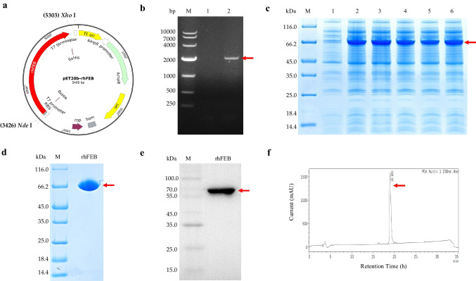
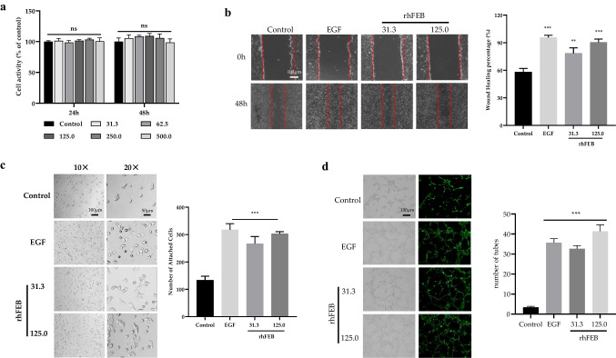
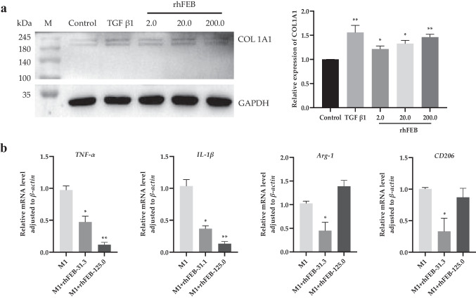
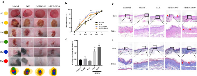
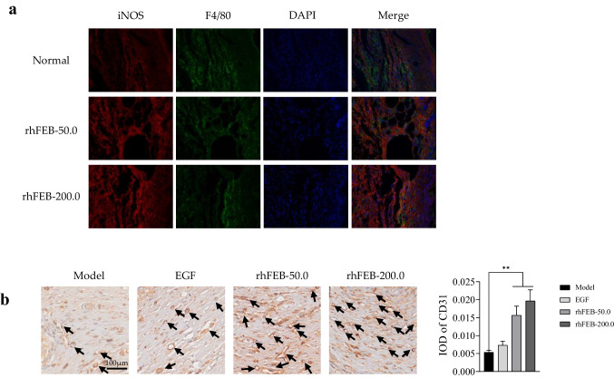
Fibronectin (FN) and collagen are vital components of the extracellular matrix (ECM). These proteins are essential for tissue formation and cell alignment during the wound healing stage. In particular, FN interacts with collagens to activate various intracellular signaling pathways to maintain ECM stability. A novel recombinant extra domain-B fibronectin (EDB-FN)-COL3A1 fusion protein (rhFEB) was designed to mimic the ECM to promote chronic and refractory skin ulcer wound healing. rhFEB significantly enhanced cell adhesion and migration, vascular ring formation, and the production of new collagen I (COL1A1) in vitro. rhFEB decreased M1 macrophages and further modulated the wound microenvironment, which was confirmed by the treatment of db/db mice with rhFEB. Accelerated wound healing was shown during the initial stages in rhFEB-treated db/db mice, as was enhanced follicle regeneration, re-epithelialization, collagen deposition, granulation, inflammation, and angiogenesis. The wound chronicity of diabetic foot ulcers (DFUs) remains the main challenge in current and future treatment. rhFEB may be a candidate molecule for regulating M1 macrophages during DFU healing. KEY POINTS: • A recombinant protein EDB-FN-collagen III (rhFEB) was highly expressed in Escherichia coli • rhFEB protein induces COL1A1 secretion in human skin fibroblasts • rhFEB protein accelerates diabetic wound healing.
Keywords: M1 macrophages; Wound healing; db/db mice; rhFEB.
© 2024. The Author(s).
Conflict of interest statement
The authors declare no competing interests.
Figures
Similar articles
-
Neurotensin-loaded collagen dressings reduce inflammation and improve wound healing in diabetic mice.Biochim Biophys Acta. 2014 Jan;1842(1):32-43. doi: 10.1016/j.bbadis.2013.10.009. Epub 2013 Oct 23. Biochim Biophys Acta. 2014. PMID: 24161538
-
HoxD3 accelerates wound healing in diabetic mice.Am J Pathol. 2003 Dec;163(6):2421-31. doi: 10.1016/S0002-9440(10)63597-3. Am J Pathol. 2003. PMID: 14633614 Free PMC article.
-
Altered ECM deposition by diabetic foot ulcer-derived fibroblasts implicates fibronectin in chronic wound repair.Wound Repair Regen. 2016 Jul;24(4):630-43. doi: 10.1111/wrr.12437. Epub 2016 Jun 8. Wound Repair Regen. 2016. PMID: 27102877 Free PMC article.
-
CD44-dependent inflammation, fibrogenesis, and collagenolysis regulates extracellular matrix remodeling and tensile strength during cutaneous wound healing.Matrix Biol. 2019 Jan;75-76:314-330. doi: 10.1016/j.matbio.2018.06.004. Epub 2018 Jun 9. Matrix Biol. 2019. PMID: 29894820 Free PMC article. Review.
-
Dermal extracellular matrix molecules in skin development, homeostasis, wound regeneration and diseases.Semin Cell Dev Biol. 2022 Aug;128:137-144. doi: 10.1016/j.semcdb.2022.02.027. Epub 2022 Mar 23. Semin Cell Dev Biol. 2022. PMID: 35339360 Review.
Cited by
-
Identifying and Validating Extracellular Matrix-Related Gene CTSH in Diabetic Foot Ulcer Using Bioinformatics and Machine Learning.J Inflamm Res. 2024 Aug 30;17:5871-5887. doi: 10.2147/JIR.S467507. eCollection 2024. J Inflamm Res. 2024. PMID: 39228680 Free PMC article.
-
Decellularized Extracellular Matrices for Skin Wound Treatment.Materials (Basel). 2025 Jun 12;18(12):2752. doi: 10.3390/ma18122752. Materials (Basel). 2025. PMID: 40572885 Free PMC article. Review.
References
-
- Barrenas F, Raehtz K, Xu C, Law L, Green RR, Silvestri G, Bosinger SE, Nishida A, Li Q, Lu W, Zhang J, Thomas MJ, Chang J, Smith E, Weiss JM, Dawoud RA, Richter GH, Trichel A, Ma D, Peng X, Komorowski J, Apetrei C, Pandrea I, Gale M Jr (2019) Macrophage-associated wound healing contributes to African green monkey SIV pathogenesis control. Nat Commun 10(1):5101. 10.1038/s41467-019-12987-9 - PMC - PubMed
-
- Bauer SM, Bauer RJ, Velazquez OC (2005) Angiogenesis, vasculogenesis, and induction of healing in chronic wounds. Vasc Endovasc Surg 39(4):293–306. 10.1177/153857440503900401 - PubMed
-
- Campos AC, Groth AK, Branco AB (2008) Assessment and nutritional aspects of wound healing. Curr Opin Clin Nutr 11(3):281–288. 10.1097/MCO.0b013e3282fbd35a - PubMed
MeSH terms
Substances
LinkOut - more resources
Full Text Sources
Miscellaneous
