

Comment
doi: 10.1093/jleuko/qiae005.
When is a Kupffer cell not a Kupffer cell? Novel insight into macrophage fate and function in hepatic fibrosis
Affiliations
- PMID: 38285520
- PMCID: PMC12020053
- DOI: 10.1093/jleuko/qiae005
Item in Clipboard
Comment
When is a Kupffer cell not a Kupffer cell? Novel insight into macrophage fate and function in hepatic fibrosis
J Leukoc Biol.
.
No abstract available
Keywords: fibrosis; liver disease; phagocytosis.
Conflict of interest statement
Conflicts of interest. The author declares that he has no known competing financial interests or personal relationships that could have appeared to influence the work reported in this article.
Figures
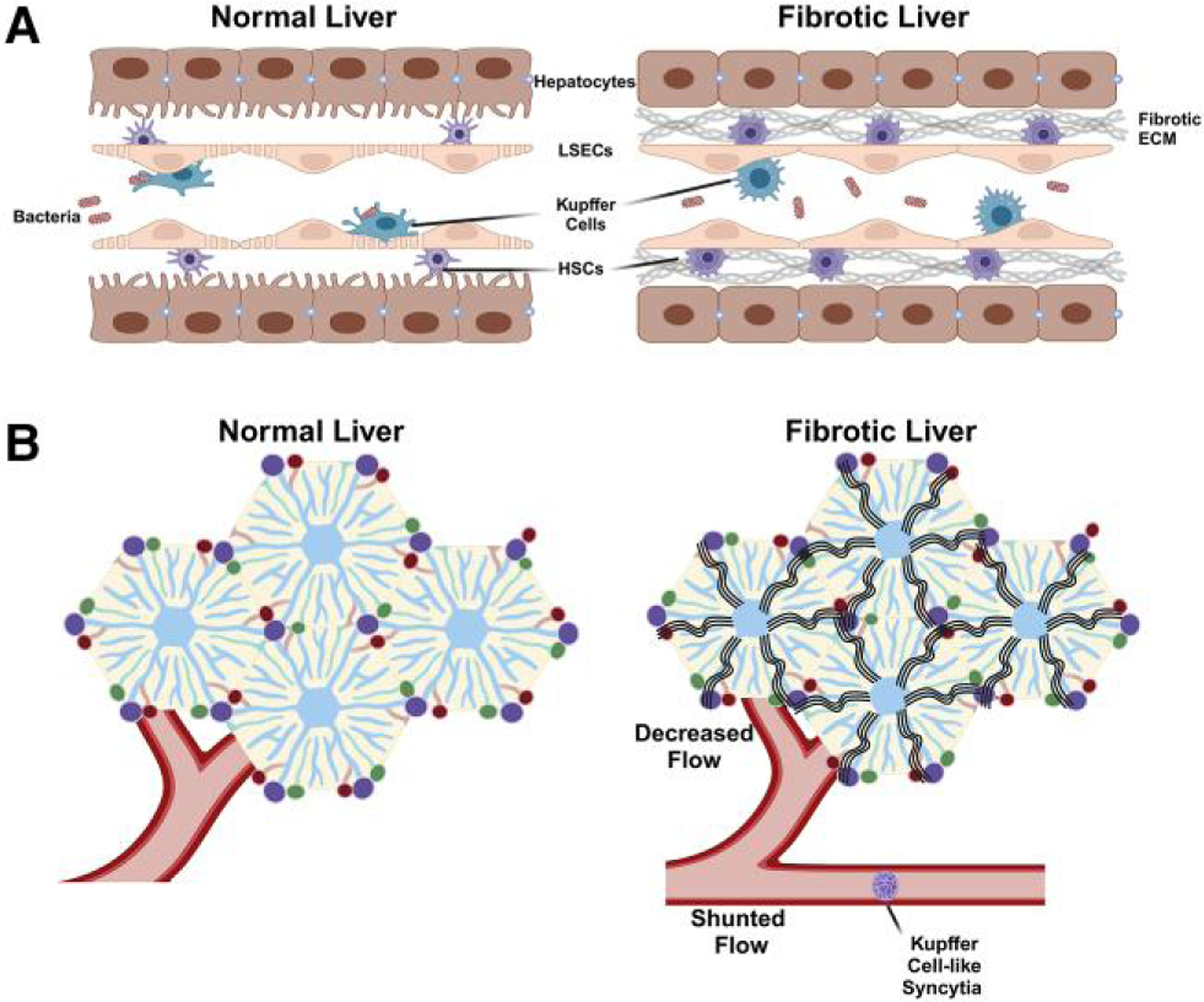
Functional adaptation to hepatic fibrosis. (A) Under normal conditions, Kupffer cells readily phagocytose xenobiotics (e.g. bacteria) that flow through the hepatic sinusoidal. This phagocytic phenotype is hypothesized to be maintained via contact with other hepatic cells, such as hepatocytes and hepatic stellate cells (HSCs). Exchange of signals between these cells is supported by the normally fenestrated liver sinusoidal endothelial cells (LSECs). During fibrosis, HSCs become activated, and extracellular matrix (ECM) into the interstitial space and endothelial cells lose their fenestrae (i.e. capillarization). Kupffer cell phagocytic function is diminished in fibrotic livers, partially via diminished cell-cell communication with hepatocytes and/or HSCs. (B) In addition to diminished phagocytic function (see panel A), fibrosis increases vascular resistance and diminishes blood flow to the hepatic lobules. The increase in vascular resistance leads to the formation of shunts that bypass the hepatic lobules. The loss of phagocytic function by Kupffer cells is partially compensated by Kupffer cell–like syncytia that form in these shunted vessels. Created with BioRender.com .
Comment on
-
Kupffer cell-like syncytia replenish resident macrophage function in the fibrotic liver.Science. 2023 Sep 8;381(6662):eabq5202. doi: 10.1126/science.abq5202. Epub 2023 Sep 8. Science. 2023. PMID: 37676943
References
-
- Kupffer C Ueber Sternzellen der Leber. Arch Mikroskop Anat. 1876:12(1):353–358. 10.1007/BF02933897 - DOI
Publication types
MeSH terms
Grants and funding
LinkOut - more resources
Full Text Sources
Medical
