. 2024 Feb 6;121(6):e2314309121.
doi: 10.1073/pnas.2314309121. Epub 2024 Jan 29.

A novel cysteine-rich adaptor protein is required for mucin packaging and secretory granule stability in vivo

Affiliations

A novel cysteine-rich adaptor protein is required for mucin packaging and secretory granule stability in vivo

Liping Zhang et al. Proc Natl Acad Sci U S A. .

Abstract

Mucins are large, highly glycosylated extracellular matrix proteins that line and protect epithelia of the respiratory, digestive, and urogenital tracts. Previous work has shown that mucins form large, interconnected polymeric networks that mediate their biological functions once secreted. However, how these large matrix molecules are compacted and packaged into much smaller secretory granules within cells prior to secretion is largely unknown. Here, we demonstrate that a small cysteine-rich adaptor protein is essential for proper packaging of a secretory mucin in vivo. This adaptor acts via cysteine bonding between itself and the cysteine-rich domain of the mucin. Loss of this adaptor protein disrupts mucin packaging in secretory granules, alters the mobile fraction within granules, and results in granules that are larger, more circular, and more fragile. Understanding the factors and mechanisms by which mucins and other highly glycosylated matrix proteins are properly packaged and secreted may provide insight into diseases characterized by aberrant mucin secretion.

Keywords: glycosylation; mucin; salivary glands; secretion; secretory granules.


Conflict of interest statement

Competing interests statement:The authors declare no competing interest.

Figures
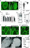
Fig. 1.
Fig. 1.
Loss of Sgs7 results in bloated secretory granules and disrupted structure of Sgs3. (A) Compared with WT SGs, RNAi to Sgs7 (Sgs7RNAi) resulted in very round, bloated secretory granules in secretory cells. Green is Sgs3-GFP packaged in secretory granules. (B) The secretory granule circularities of WT and Sgs7RNAi were quantified as described previously (24). 200 granules of each genotype were analyzed. ****P < 0.0001. (C) Expression of each Sgs gene was quantitated in WT and Sgs7RNAi SGs by qPCR. Among six Sgs genes, only Sgs7 expression was dramatically decreased in Sgs7RNAi SGs, while others were unchanged. (D) Coomassie blue staining of proteins from WT and Sgs7RNAi SGs showed a specific decrease in a protein with the predicted size of Sgs7. (E) Western blot with Sgs7 antibody further confirmed Sgs7 protein level was dramatically decreased in Sgs7RNAi SGs. (F) Transposon mutations of Sgs7 (Sgs7Bl#24539/Bl#57993) also resulted in bloated secretory granules, similar to Sgs7RNAi. Green is GFP expressed in the cytoplasm of secretory cells. (G) Expression of Sgs7 in Sgs7 mutant SGs (Sgs7Bl#24539/Bl#57993; c135>UAS-Sgs7-V5) rescued the abnormal secretory granule phenotype. (H) TEM on secretory granules of WT and Sgs7RNAi. In WT secretory granules, Sgs3 is present as highly ordered bundled filaments (red arrowhead). In Sgs7RNAi, no bundled filaments were seen (black arrowhead) but other structures seen in WT were present at the periphery. (I) Western blot probed with Sgs3 antibody demonstrated that Sgs3 protein was present at similar levels in WT and Sgs7RNAi SGs, (black arrowheads). (Scale bars are 20 µm for (A), (F), and (G) and 600 nm for (H).)
Fig. 2.
Fig. 2.
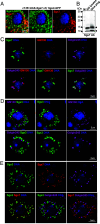
Sgs7 and Sgs3 colocalize in secretory granules. (A) Staining of SGs with the anti-V5 antibody showed that Sgs7 (red) co-localizes with truncated Sgs3-GFP transgene (green) in secretory granules. (Scale bar, 20 µm.) (B) Western blot probed with the Sgs7 antibody showed that Sgs7 protein is present in secretory cells before secretion (SG lysates) and also within the secreted material (glue plug). (C) Sgs7 (green) expressed in Drosophila S2R+ cells localized to the Golgi apparatus, as detected by the cis-Golgi marker GM130 (red) and the trans-Golgi marker Golgin245 staining (magenta). DNA staining is shown in blue. (D) Full-length Sgs3 (green) expressed in S2R+ cells formed secretory granule-like structures and did not localize to Golgi apparatus (GM130 or Golgin245, magenta). (E) When co-expressed with Sgs3 (green), Sgs7 (red) no longer localizes to the Golgi (Golgin245, magenta) but co-localized with Sgs3 in vesicle-like structures. (Scale bar, 5 µm.)
Fig. 3.
Fig. 3.
Sgs3 and Sgs7 interact through cysteine bonding. (A) Diagram of WT Sgs3 and different domain deletions used for co-expression with Sgs7. N, N-terminal domain; T-rich, threonine-rich domain; PTTTK, repetitive mucin domain; C, cysteine-rich C-terminal domain. (B) The deletions of the T-rich domain (Sgs3∆T, green) or the PTTTK domain (Sgs3∆P, green) co-localized with Sgs7 (red). Deletions that removed the C-terminal region (Sgs3∆PC or Sgs3∆C, green) resulted in loss of colocalization with Sgs7 (red). Additionally, Sgs3∆C disrupted the formation of large secretory granule-like structures. (C) Alignment of Sgs7 and Sgs3 revealed considerable homology within the cysteine-rich regions of both proteins. Cysteines are highlighted. Mutating the cysteines within the cysteine-rich domain of Sgs3 (Sgs3M, green) (D) or Sgs7 (Sgs7M, red) (E) resulted in the loss of colocalization of these proteins. Additionally, Sgs3M was unable to form granule-like structures. (F) Western blots demonstrate that Sgs3 (Sgs3-FLAG) was co-precipitated with WT Sgs7 (Sgs7-AV5) but not with the cysteine mutated Sgs7 (Sgs7M-AV5). (G) Mutated Sgs7 (Sgs7M) was unable to rescue the bloated secretory granules (Sgs7Bl#24539/Bl#57993; c135>UAS-Sgs7M-V5). Green is the GFP expressed in the cytoplasm of secretory cells. (H) AlphaFold2 was used to model complex formation between Sgs3 and Sgs7. Formation of tetramers by two Sgs7 molecules and two Sgs3 molecules. Red is intra-disulfide-bond and magenta is inter-disulfide-bond. White dotted box shows a magnified view of the intra- and inter-disulfide-bonds formed in the tetramer. There is one intra-disulfide-bond in each Sgs7 (C31-C38) and Sgs3 (C264-C271), two inter-disulfide-bonds between two Sgs7 molecules (two C41-C65 bonds) and Sgs3 (two C274-C298 bonds), and four inter-disulfide-bonds between two Sgs7 and two Sgs3 molecules (two Sgs7C67-Sgs3C281 bonds and two Sgs7C48-Sgs3C300 bonds). See also Movie S1. (Scale bars are 5 µm for (B), (D), and (E) and 20 µm for (G).)
Fig. 4.
Fig. 4.
Sgs7RNAi results in altered intragranular mobility and fragile secretory granules. (A) FRAP analysis of secretory granules of live SGs from WT or Sgs7RNAi larvae. A small circular region of interest (ROI) in a secretory granule containing Sgs3-GFP (shown in white) was defined, and the fluorescence intensity change in ROI before and after the bleach (Fpre-bleach and Fpost-bleach) was monitored over time. (B) Fluorescence intensity values of 20 secretory granules from WT or Sgs7RNAi were plotted and used to generate values for recovery half times (t1/2) (C) and mobile fraction (Mf) (D), which were calculated based on the equations described in Snapp et al. (35). (E) AFM on secretory granules from WT or Sgs7RNAi larvae to calculate elastic modulus of granule contents. The beginning and end time of incubation of WT (F) and Sgs7RNAi (G) SGs in a hypotonic solution. See also Movies S2 and S3. Note the dark regions, indicating ruptured secretory granules, in the end time point of Sgs7RNAi. (Scale bars are 5 µm for (A) and 50 µm for (F) and (G).)

Similar articles

Cited by

References

    1. Raote I., Malhotra V., Protein transport by vesicles and tunnels. J. Cell Biol. 218, 737–739 (2019). - PMC - PubMed
    1. Valentijn K. M., Sadler J. E., Valentijn J. A., Voorberg J., Eikenboom J., Functional architecture of Weibel-Palade bodies. Blood 117, 5033–5043 (2011). - PMC - PubMed
    1. Ermund A., Trillo-Muyo S., Hansson G. C., Assembly, release, and transport of airway mucins in pigs and humans. Ann. Am. Thorac. Soc. 15, S159–S163 (2018). - PMC - PubMed
    1. Johansson M. E., et al. , Composition and functional role of the mucus layers in the intestine. Cell Mol. Life Sci. 68, 3635–3641 (2011). - PMC - PubMed
    1. Ambort D., et al. , Calcium and pH-dependent packing and release of the gel-forming MUC2 mucin. Proc. Natl. Acad. Sci. U.S.A. 109, 5645–5650 (2012). - PMC - PubMed

LinkOut - more resources