Review
. 2024 Feb;45(1):3-20.
doi: 10.1055/s-0043-1777259. Epub 2024 Jan 29.

Aspergillus and the Lung

Affiliations
Review

Aspergillus and the Lung

Iris Janssens et al. Semin Respir Crit Care Med. 2024 Feb.

Abstract

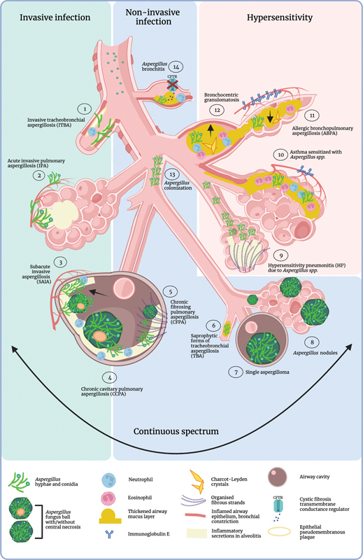
The filamentous fungus Aspergillus causes a wide spectrum of diseases in the human lung, with Aspergillus fumigatus being the most pathogenic and allergenic subspecies. The broad range of clinical syndromes that can develop from the presence of Aspergillus in the respiratory tract is determined by the interaction between host and pathogen. In this review, an oversight of the different clinical entities of pulmonary aspergillosis is given, categorized by their main pathophysiological mechanisms. The underlying immune processes are discussed, and the main clinical, radiological, biochemical, microbiological, and histopathological findings are summarized.


Conflict of interest statement

None declared. E.V.B. is chair of the Chronic Pulmonary Aspergillosis Network (CPAnet).

Figures

Fig. 1
Fig. 1
Clinical spectrum of pulmonary aspergillosis. The clinical spectrum of pulmonary aspergillosis is divided into three categories: invasive infection ( left panel ), noninvasive infection ( middle panel ), and manifestations of hypersensitivity ( right panel ). The different clinical entities that can develop from the presence of Aspergillus are numbered 1 to 14, accompanied by an illustration of their main pathophysiological characteristics. (1) Invasive tracheobronchial aspergillosis (ITBA) is described in an ulcerative form and a pseudomembranous form; here, the pseudomembranous form is depicted with pseudomembranous inflammatory ulcerative plaques in a proximal airway with invasiveness in the enrobing tissue. (2) Acute invasive pulmonary aspergillosis (IPA), depicted here in a neutropenic environment with fungal hyphae invading the alveolar capillary, pointing out the tissue- and possible angio-invasiveness of the disease. (3) Subacute invasive aspergillosis (SAIA), a disease entity with characteristics of both IPA and chronic cavitary pulmonary aspergillosis. The rapid progression of cavitation in this disease is indicated with the arrow. Tissue invasiveness is often described. (4) Chronic cavitary pulmonary aspergillosis (CCPA) is characterized by one or more cavities with or without a thickened wall. The cavities can be filled with solid or fluid material and a fungal ball can be present. Local tissue invasion may occur depending on the host immune status. (5) Chronic fibrosing pulmonary aspergillosis (CFPA) is considered a fibrotic end-stage of CCPA. (6) Saprophytic forms of tracheobronchial aspergillosis (TBA) are bronchial aspergillosis, endobronchial aspergillosis, and mucoid impaction. The fungal hyphae are restricted to the airway lumen. (7) A single aspergilloma does not induce a clinical inflammatory response, neither does an Aspergillus nodule (8). (9) Hypersensitivity pneumonitis (HP) due to Aspergillus spp. can present with or without fibrosis. (10) Asthma sensitized by Aspergillus spp. involves airway constriction, mucus production and a T helper 2 (Th2) immune response with eosinophilia and immunoglobulin E (IgE) production. (11) Allergic bronchopulmonary aspergillosis (ABPA) involves Th2 inflammation with eosinophilia, Charcot–Leyden crystals, and overzealous IgE production which results in mucus plugging, airway constriction and congestion, central bronchiectasis (indicated by the black arrow pointing upward), and atelectasis (indicated by the black arrow pointing downwards). (12) Bronchocentric granulomatosis can present as ABPA that is confined to the bronchi. (13) Uncomplicated airway colonization with Aspergillus spp. (14) Aspergillus bronchitis is described in cystic fibrosis (CF) and non-CF bronchiectasis. Bronchiectasis and sticky airway mucus in CF is a result from the absence or malfunctioning of the ion transporter cystic fibrosis transmembrane conductance regulator (CFTR). These 14 different disease entities are not strictly delineable and make part of a continuous clinical spectrum. One form of clinical disease may evolve into another over time, depending on the degree of immune compromise or hyperresponsiveness of the host. Each of these different disease presentations can evolve into one another irrespective of the initial immunopathogenesis. Moreover, different disease entities may exist at the same time. This figure was created with BioRender.
Fig. 2
Fig. 2
Pathogenesis of acute angio-invasive and tissue-invasive pulmonary aspergillosis (IPA).Aspergillus spores are inhaled and, in the right circumstances, shed their hydrophobic rodlet layer and germinate into hyphae. In the absence of neutrophils (1), caused by the underlying disease or by the treatment regimen, neutrophil phagocytosis, neutrophil extracellular trap formation (NETosis) and neutrophil signaling is impaired, and fungal hyphae protrude into the lung tissue and the blood vessels, causing disseminated disease. In nonneutropenic patients, IPA can develop when another important immune defect is present, for example, lack of CD4 T cells in acquired immunodeficiency syndrome (AIDS) (2) or a severe acute respiratory syndrome coronavirus type 2 (SARS-CoV-2) or influenzavirus infection (3). However, angio-invasion is usually not observed without profound and prolonged neutropenia. Abbreviations: AIDS, acquired immunodeficiency syndrome; IPA, invasive pulmonary aspergillosis; NETosis, neutrophil extracellular trap formation; SARS-CoV-2, severe acute respiratory syndrome coronavirus type 2. This figure was created with BioRender.
Fig. 3
Fig. 3
Ulcerative invasive tracheobronchial aspergillosis (ITBA). Endoscopic image of ulcerative ITBA in a 43-year-old male that was treated for hairy cell leukemia. A sharply delineated ulcerative plaque covers the carina and a part of the left main stem bronchus. A. fumigatus was isolated from a cultured biopsy. Image courtesy of Thomas Malfait, MD.
Fig. 4
Fig. 4
Mucusplugging in the right lower lobe of a patient with cystic fibrosis and allergic bronchopulmonary aspergillosis (ABPA). Panel A shows a multidetector computed tomography (MDCT) section with an unplugged bronchus in the right lower lobe (indicated by the arrow), whereas in panel B, this very airway is plugged, causing lung parenchyma consolidation and ground-glass opacification along the more peripheral tract of the bronchus (indicated by the arrow). In panel C, an endoscopic image is shown with complete obliteration of the lateral segment of the right lower lobe by a mucus plug. In panel D, this plug is removed, revealing inflammatory bronchial epithelium. In panel E, a photograph of the removed mucus plug is shown, its total length is 4 centimeter. In panel F, a Rapid-Chrome Kwik–Diff staining from this sputum plug cytospin is shown. An eosinophil is indicated with the black arrow and two Charcot–Leyden crystals are indicated with the blue arrows. Endoscopic images courtesy of Yannick Vande Weygaerde, MD.
Fig. 5
Fig. 5
The pathogenesis of fungal sensitized asthma and allergic bronchopulmonary aspergillosis (ABPA). Panel A shows how fungal allergens act as proteases that directly affect the respiratory epithelium: (1) in murine models, alkaline serine protease 1 (Alp1) degrades club cell junctions and the underlying extracellular matrix. This leads to smooth muscle contraction and airway hyperresponsiveness. (2) The junctional damage activates the mechanosensitive calcium channel TRPV4, which in turn causes calcium influx, calcineurin signaling and induction of a Th2 immunological response. (3) Aspergillus proteases cleave IL-33 into a highly active form that activates ILC2s to produce type 2 cytokines and drive IL-5-dependent eosinophil recruitment to the lungs. Panel B shows that Th2 immunity and mucus plug formation are the underlying immunopathogenesis of allergic fungal airway disease with ABPA as the most advanced presentation. In ABPA, Charcot–Leyden crystals (CLCs) tie the components of the plug together in a complex mesh. See text for a detailed description. Abbreviations: Alp1, alkaline serine protease 1; APC, antigen-presenting cell; BC, B cell; CC, club cell; ECM, extracellular matrix; eDNA, extracellular DNA; EET, eosinophil extracellular trap; Eo, eosinophil; EP, epithelium; EPO, eosinophil peroxidase; Gal-10: galectin-10; GC, goblet cell; H2O2, hydrogen peroxide; Ig, immunoglobulin; IL, interleukin; ILC2, innate like cell type 2; MC, mast cell; MPO, myeloperoxidase; NE, neutrophil elastase; NET, neutrophil extracellular trap; Neutro, neutrophil; PC, plasma cell; Th, T helper; TRPV4, transient receptor potential cation channel subfamily V member 4. This figure was created with BioRender.
Fig. 6
Fig. 6
Bronchocentric granulomatosis. Endoscopic image of bronchocentric granulomatosis in an 82-year-old female with a history of pulmonary tuberculosis. The granuloma is calcified and overgrown with Aspergillus hyphae. Image courtesy of Thomas Malfait, MD and Philippe Rogiers, MD.

References

    1. Patterson T F. Elsevier; 2019. Aspergillus Species.
    1. Griffiths J S, Camilli G, Kotowicz N K, Ho J, Richardson J P, Naglik J R. Role for IL-1 family cytokines in fungal infections. Front Microbiol. 2021;12:633047. - PMC - PubMed
    1. Kosmidis C, Denning D W. The clinical spectrum of pulmonary aspergillosis. Thorax. 2015;70(03):270–277. - PubMed
    1. Brown G D, Denning D W, Gow N AR, Levitz S M, Netea M G, White T C. Hidden killers: human fungal infections. Sci Transl Med. 2012;4(165):165rv13. - PubMed
    1. Ainsworth G C. Cambridge University Press;; 1976. Introduction to the History of Mycology.