. 2024 May 20;79(6):524-537.
doi: 10.1136/thorax-2022-219958.

COPD basal cells are primed towards secretory to multiciliated cell imbalance driving increased resilience to environmental stressors

Affiliations

COPD basal cells are primed towards secretory to multiciliated cell imbalance driving increased resilience to environmental stressors

Mircea Gabriel Stoleriu et al. Thorax. .

Abstract

Introduction: Environmental pollutants injure the mucociliary elevator, thereby provoking disease progression in chronic obstructive pulmonary disease (COPD). Epithelial resilience mechanisms to environmental nanoparticles in health and disease are poorly characterised.

Methods: We delineated the impact of prevalent pollutants such as carbon and zinc oxide nanoparticles, on cellular function and progeny in primary human bronchial epithelial cells (pHBECs) from end-stage COPD (COPD-IV, n=4), early disease (COPD-II, n=3) and pulmonary healthy individuals (n=4). After nanoparticle exposure of pHBECs at air-liquid interface, cell cultures were characterised by functional assays, transcriptome and protein analysis, complemented by single-cell analysis in serial samples of pHBEC cultures focusing on basal cell differentiation.

Results: COPD-IV was characterised by a prosecretory phenotype (twofold increase in MUC5AC+) at the expense of the multiciliated epithelium (threefold reduction in Ac-Tub+), resulting in an increased resilience towards particle-induced cell damage (fivefold reduction in transepithelial electrical resistance), as exemplified by environmentally abundant doses of zinc oxide nanoparticles. Exposure of COPD-II cultures to cigarette smoke extract provoked the COPD-IV characteristic, prosecretory phenotype. Time-resolved single-cell transcriptomics revealed an underlying COPD-IV unique basal cell state characterised by a twofold increase in KRT5+ (P=0.018) and LAMB3+ (P=0.050) expression, as well as a significant activation of Wnt-specific (P=0.014) and Notch-specific (P=0.021) genes, especially in precursors of suprabasal and secretory cells.

Conclusion: We identified COPD stage-specific gene alterations in basal cells that affect the cellular composition of the bronchial elevator and may control disease-specific epithelial resilience mechanisms in response to environmental nanoparticles. The identified phenomena likely inform treatment and prevention strategies.

Keywords: Airway Epithelium; COPD Pathology; COPD exacerbations mechanisms; Occupational Lung Disease; Thoracic Surgery.


Conflict of interest statement

Competing interests: None declared.

Figures

Figure 1
Figure 1
Increased resilience of COPD stage-IV derived pHBEC towards airborne toxin exposure associated with altered secretory to multiciliated cell balance at ALI. (A) Schematic representation of the experimental design demonstrating exposure of long-term ALI cultured pHBEC to environmentally relevant aerosolised cytotoxic (ZnO) and proinflammatory (CNP, LPS) nanoparticles (NP). (B) Analysis of membrane (left, lactate dehydrogenase (LDH) assay) and barrier (middle, transepithelial electrical resistance (TEER)) integrity and metabolic cell viability (right, tetrazolium salt assay (WST-1)) of pHBECs from non-CLD, COPD-II and COPD-IV patients 24 hours after NP exposure, showing a diminished susceptibility to ZnO exposure induced cellular damage indicated by reduced LDH release and cell barrier integrity loss as well as a preserved metabolic cell activity in COPD-IV cultures when compared with non-CLD and COPD-II cultures. Results are derived from n=4 non-CLD, n=3 COPD-II and n=4 COPD-IV pHBECs cultures and presented as median and IQR. (C, D) Exemplary images (three-dimensional microscopy) and quantification of confocal IF images of NP exposed pHBEC ALI cultures after antibody staining for secretory (MU5AC+ and CC10+) and multiciliated cells (Ac-Tub+), showing an unchanged number of multiciliated cells in exposed non-CLD pHBECs, in contrast to a decrease in multiciliated cell number in COPD-II cultures and an unchanged number of secretory cells in both non-CLD and COPD-II pHBECs at ALId28. Results are derived from n=3 non-CLD, n=3 COPD-II and n=3 COPD-IV pHBEC cultures with IF quantification performed in 3–5 representative fields per transwell per condition (magnification ×63). Results are presented as median and IQR. Data information: Kruskal Wallis test with Bonferroni post hoc analysis was used for group comparisons. Results presented as median and IQR. Sham controls (saline solution 0.9%), LPS 1 mg/mL, CNP 10.8cm2/cm2, ZnO low dose 0.14 cm2/cm2 and ZnO high dose 3.19 cm2/cm2 at ALId30±2 after air-lift. Scale bar: 50 µm. ALI, air–liquid interface; CNP, carbon nanoparticle; COPD, chronic obstructive pulmonary disease; LPS, lipopolysaccharide; pHBEC, primary human bronchial epithelial cell.
Figure 2
Figure 2
Healthy and diseased ALI cultures reveal shared gene expression and pathway regulation on LPS and ZnO exposure. (A) Pathway enrichment analysis from bulk transcriptome of non-CLD controls and COPD-IV ALI cultures, showing the presence of cilium regulation mechanisms in COPD-IV cultures on low-dose ZnO exposure. (B) Upregulation of innate defence mechanisms in both healthy and diseased ALI cultures in response to LPS exposure. (C) The heatmap illustrates the top 25 significantly upregulated genes from bulk transcriptome analysis of the LPS-exposed non-CLD and COPD-IV ALI cultures. Gene expression indicates a comparable response with regard to nanoparticle clearance. Genes with adjusted p values<0.1 are marked with an asterisk. Data information: Regulated genes were filtered by using raw p values<0.05 and FC (fold change) ≥1.3. log2FC are shown. Bulk transcriptome analysis was derived from n=3 non-CLD and n=3 COPD-IV pHBEC cultures. ALI, air–liquid interface; CLD, chronic lung disease; COPD, chronic obstructive pulmonary disease; LPS, lipopolysaccharide; pHBEC, primary human bronchial epithelial cell.
Figure 3
Figure 3
Altered cell differentiation in COPD stage-IV derived pHBEC at ALI resulted from basal cells imbalance. (A) Analysis of epithelial barrier integrity via transepithelial electrical resistance (TEER) of pHBEC isolated from non-CLD, COPD-II (native and+CSE) and COPD-IV patients, showing slightly reduced TEER values in COPD-II CSE when compared with the untreated COPD-II cultures. Results are derived from n=4 non-CLD, n=3 COPD-II, n=3 COPD-II CSE and n=4 COPD-IV pHBEC cultures. (B) Quantification of confocal three-dimensional IF microscopy of non-CLD, COPD-II, COPD-II CSE and COPD-IV derived pHBECs at ALId28 demonstrating a decrease in the number of multiciliated (Ac-Tub+) of COPD-II treated cultures vs untreated COPD-II and non-CLD cultures at ALId28. Secretory cell number remained almost unchanged on CSE exposure. The remaining cells from the total number of cells at ALId28 reflect the number of basal and suprabasal cells on the end-differentiated ALI cultures. Results are derived from n=3 non-CLD, n=3 COPD-II, n=3 COPD-II CSE and n=3 COPD-IV pHBEC cultures with IF quantification performed in 3–5 representative fields per condition. (C) IF microscopy of confocal IF images in sections of native bronchial tissue samples from n=2 COPD-IV and n=2 non-CLD patients (magnification ×20). (D) Quantification of confocal IF images in sections of native bronchial tissue samples from COPD-IV and non-CLD patients demonstrating the reduced presence of multiciliated (Ac-Tub+) cells and the dominant presence of MU5AC+ and CC10+ epithelial cells in COPD-IV cultures. Results are derived from n=2 non-CLD and n=2 COPD-IV pHBEC cultures with IF quantification performed in 3–5 representative fields per condition. (E) Optical microscopy of paraffin-embedded bronchial sections derived from n=2 COPD-IV and n=2 non-CLD patients stained with H&E as well as E-cadherin, showing an irregular and thicker basement membrane, more secretory cells and less multiciliated cells in COPD-IV in comparison to the non-CLD samples (magnification ×20). (F) Ciliary beating frequency (CBF, Hz) in non-CLD, COPD-II and COPD-IV derived pHBEC cultures at ALId28+1, at baseline level, demonstrating an altered, biphasic CBF pattern in COPD-IV with (aberrantly) slow and fast beating multiciliated cells and a physiological CBF between 7 and 16 Hz in non-CLD and COPD-II cultures. Results are derived from n=2 non-CLD, n=2 COPD-II and n=2 COPD-IV cultures at baseline level. Data information: Results presented as median and IQR. Comparative analysis for IF quantification of native bronchial tissue samples was performed by Wilcoxon test; and for CBF by Kolmogorov-Smirnov test adjusted by Benjamini-Hochberg test for multiple testing correction. Scale bar: 10 µm/20 µm. ALI, air–liquid interface; CLD, chronic lung disease; COPD, chronic obstructive pulmonary disease; CSE, cigarette smoke extract; pHBEC, primary human bronchial epithelial cell.
Figure 4
Figure 4
Longitudinal single-cell RNA-seq reveals altered differentiation dynamics in COPD-IV derived ALI cultures (n=2) in comparison to the non-CLD ALI cultures (n=2). (A) Schematic representation of the experimental design: ALI culture of pHBECs derived from n=2 non-CLD and n=2 COPD-IV patients followed by serial sampling and single-cell RNA-seq (Drop-seq) analysis on ALId0, 3, 5, 7, 14, 21, 28. (B, C) The indicated colour codes on UMAP and diffusion map (DC) embeddings show time points of sampling and cell type annotations for non-CLD control pHBEC B) and COPD-IV C) cultures. (D, E) The matchScore coefficients indicate the transcriptional similarity of cell type annotations in the pHBEC cultures to a in vivo single-cell RNA-seq reference dataset obtained from healthy individuals. (F) The stacked bar graph shows changes in relative proportions of cell types generated in the pHBEC cultures at the indicated time points. (G) The bar graphs show the percentage of proliferating cells (MKI67+/TOP2A+ ) in the indicated experimental conditions and cell types. Data information: Results are derived from n=2 non-CLD and n=2 COPD-IV immediately processed pHBEC cultures following dispase dissociation protocol. Wilcoxon rank-sum test, Bonferroni corrected. ALI, air–liquid interface; CLD, chronic lung disease; COPD, chronic obstructive pulmonary disease; pHBEC, primary human bronchial epithelial cell.
Figure 5
Figure 5
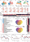
An altered basal cell state in ALI COPD-IV pHBECs propagates into its differentiated progeny. (A, B) UMAP embedding overlaid with sample identifier and cell type annotation for starting cells at ALId0 A) and endpoint of the differentiation at ALId28 B) for patients from both health conditions. (C) Feature plots of common marker genes of airway epithelial populations to confirm cell type labels at ALId28. Selected genes distinguishing the two basal populations (basal_1 for COPD-IV or basal_2 for non-CLD cultures). (D) Top 30 upregulated genes in either basal_1 or basal_2 population derived from differential tests between basal_1 and basal_2 cells. Genes that were significantly regulated on both ALId0 and 28 cells are shown. (E) Significantly enriched terms in the basal_1 and basal_2 signature (false discovery rate/FDR<0.05). (F, G) Venn-diagrams summarising the number of overlapping genes that were upregulated (F) or downregulated (G) at ALId28 COPD-IV cultures per cell type compared with non-CLD cultures. (H) Heatmap reflecting top 30 consistently upregulated and downregulated genes in COPD-IV in all cell populations at ALId28. Results are based on differential tests between COPD-IV and non-CLD for each cell type separately. (I–K) Boxplots showing the enrichment of gene signatures for the GO Terms ‘cytosolic ribosome’ (I), ‘Notch binding’ (J) and the UniProt keyword ‘Wnt signalling pathway’ (K) split by health status. Data information: Results are derived from n=2 non-CLD and n=2 COPD-IV immediately processed pHBEC cultures following dispase dissociation protocol. Wilcoxon rank-sum test, Bonferroni corrected. ALI, air–liquid interface; CLD, chronic lung disease; COPD, chronic obstructive pulmonary disease; pHBEC, primary human bronchial epithelial cell.
Figure 6
Figure 6
Altered secretory to multiciliated cell balance in COPD-IV-derived pHBECs is associated with increased Notch pathway activity. (A, B) Boxplots showing the enrichment of the multiciliated (A) and secretory (B) cell type signature split by health status. Time points are pooled for all cell types, and progression of score over time is highlighted in relevant cell populations as indicated. (C) Gene kinetics of cell type markers for basal cells, multiciliated cells and secretory cells over the course of the experiment revealing disease-induced shifts propagated over time. (D, E) Boxplots summarising enrichment scoring results based on genes associated with the GO term ‘Notch binding’ over time, shown for only multiciliated (D) and secretory cells (E) separately. Data information: Results are derived from n=2 non-CLD and n=2 COPD-IV immediately processed pHBEC cultures following dispase dissociation protocol. Wilcoxon rank-sum test, Bonferroni corrected. CLD, chronic lung disease; COPD, chronic obstructive pulmonary disease; pHBEC, primary human bronchial epithelial cell.

References

    1. Kido T, Tamagawa E, Bai N, et al. . Particulate matter induces translocation of IL-6 from the lung to the systemic circulation. Am J Respir Cell Mol Biol 2011;44:197–204. 10.1165/rcmb.2009-0427OC - DOI - PubMed
    1. Xu X, Ha SU, Basnet R. A review of epidemiological research on adverse neurological effects of exposure to ambient air pollution. Front Public Health 2016;4:157. 10.3389/fpubh.2016.00157 - DOI - PMC - PubMed
    1. Greenberg MI, Vearrier D. Metal fume fever and polymer fume fever. Clin Toxicol (Phila) 2015;53:195–203. 10.3109/15563650.2015.1013548 - DOI - PubMed
    1. Maier KL, Alessandrini F, Beck-Speier I, et al. . Health effects of ambient particulate matter--biological mechanisms and inflammatory responses to in vitro and in vivo particle exposures. Inhal Toxicol 2008;20:319–37. 10.1080/08958370701866313 - DOI - PubMed
    1. Hansel NN, McCormack MC, Kim V. The effects of air pollution and temperature on COPD. COPD 2016;13:372–9. 10.3109/15412555.2015.1089846 - DOI - PMC - PubMed

Publication types