. 2024 May;45(5):926-944.
doi: 10.1038/s41401-023-01223-2. Epub 2024 Jan 29.

Desloratadine alleviates ALS-like pathology in hSOD1G93A mice via targeting 5HTR2A on activated spinal astrocytes

Affiliations

Desloratadine alleviates ALS-like pathology in hSOD1G93A mice via targeting 5HTR2A on activated spinal astrocytes

Jian Lu et al. Acta Pharmacol Sin. 2024 May.

Abstract

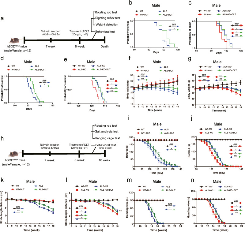
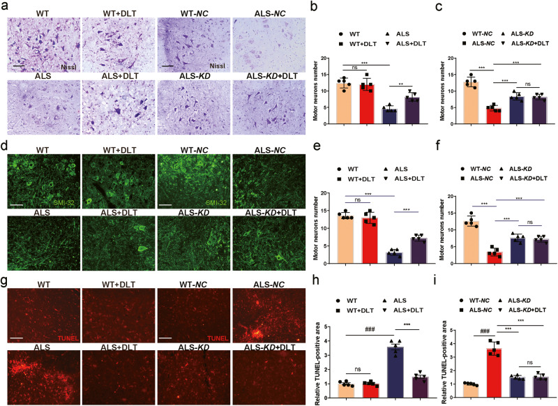
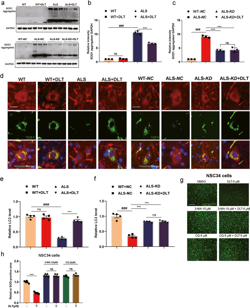
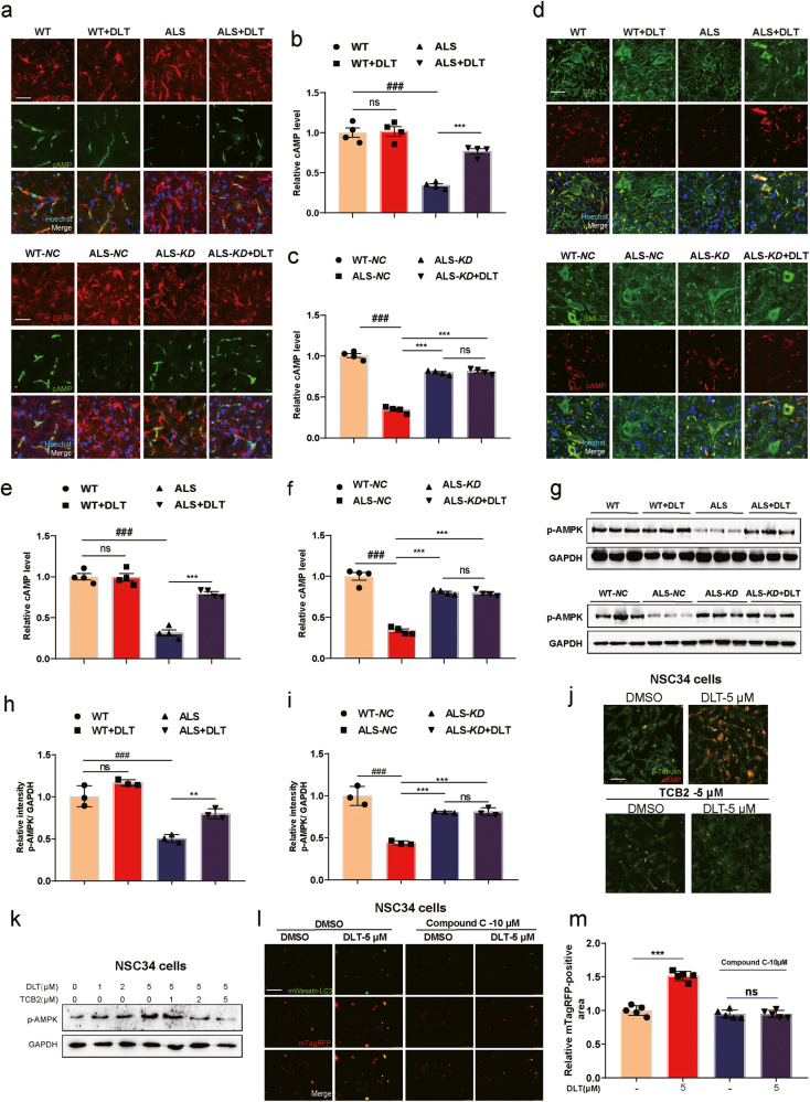
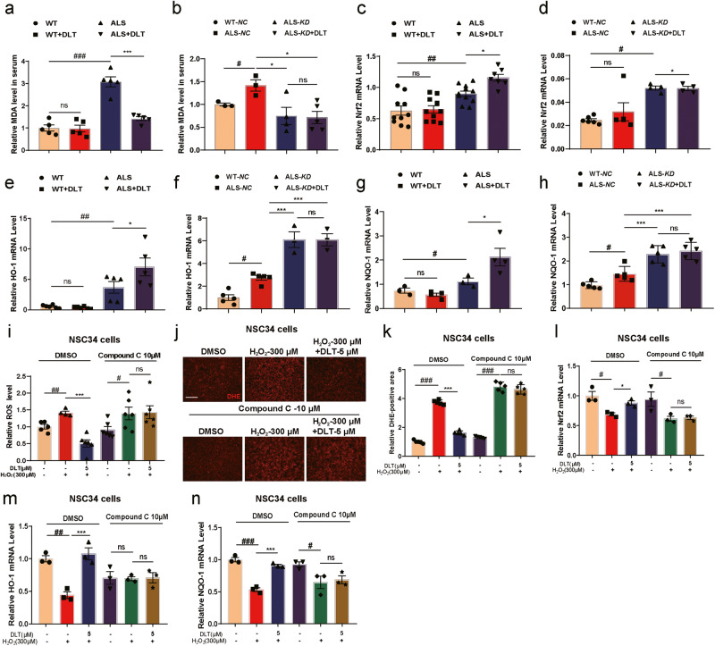
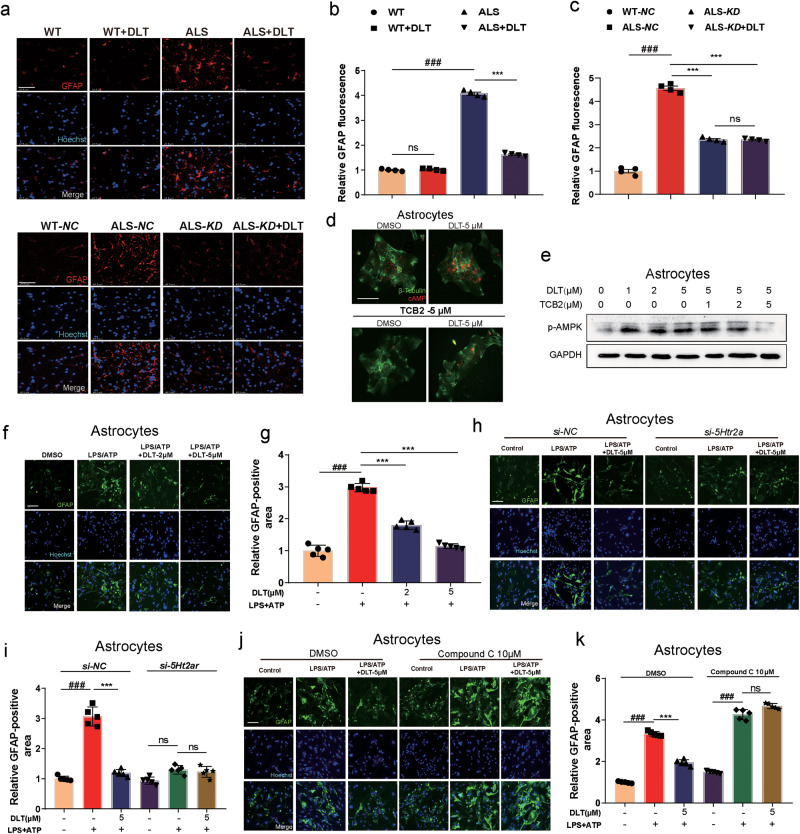
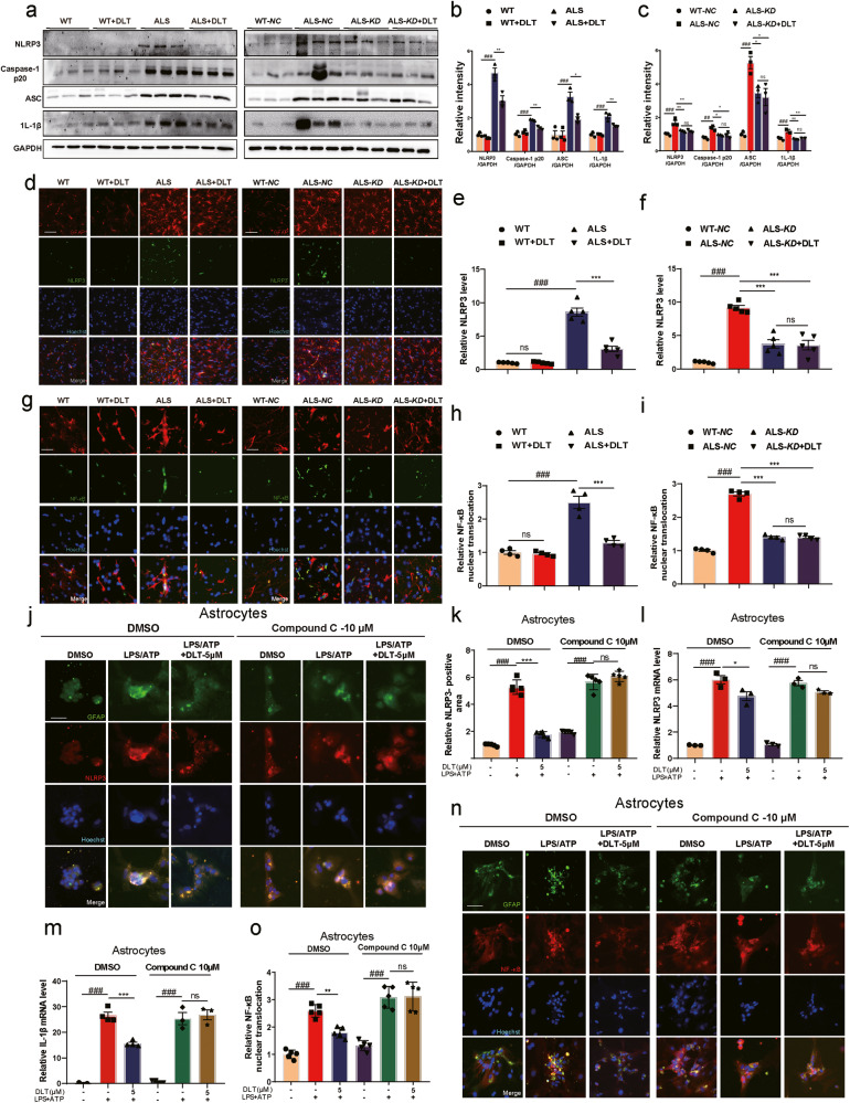
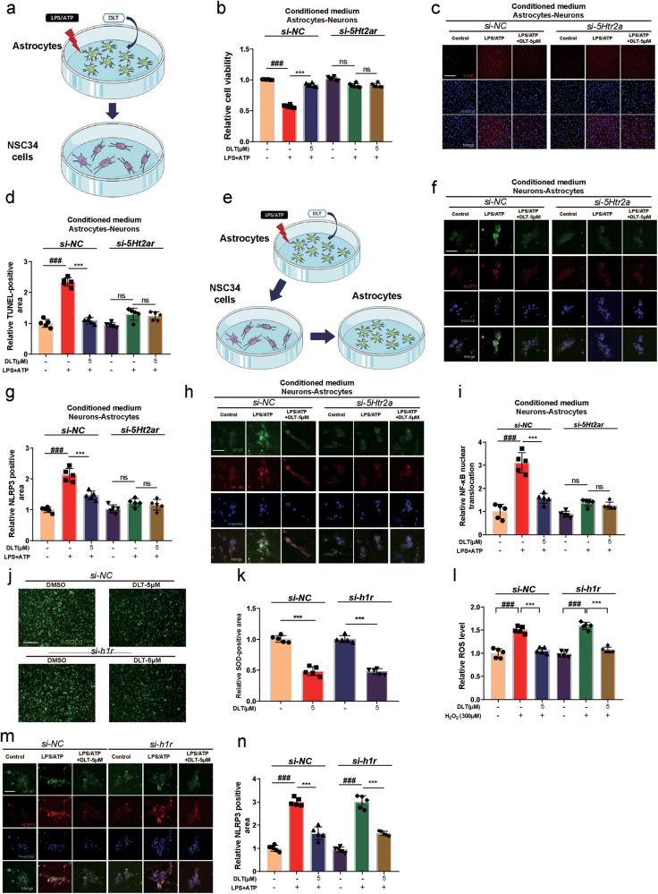
Amyotrophic lateral sclerosis (ALS) is a fatal neurodegenerative disease with progressive loss of motor neurons in the spinal cord, cerebral cortex and brain stem. ALS is characterized by gradual muscle atrophy and dyskinesia. The limited knowledge on the pathology of ALS has impeded the development of therapeutics for the disease. Previous studies have shown that autophagy and astrocyte-mediated neuroinflammation are involved in the pathogenesis of ALS, while 5HTR2A participates in the early stage of astrocyte activation, and 5HTR2A antagonism may suppress astrocyte activation. In this study, we evaluated the therapeutic effects of desloratadine (DLT), a selective 5HTR2A antagonist, in human SOD1G93A (hSOD1G93A) ALS model mice, and elucidated the underlying mechanisms. HSOD1G93A mice were administered DLT (20 mg·kg-1·d-1, i.g.) from the age of 8 weeks for 10 weeks or until death. ALS onset time and lifespan were determined using rotarod and righting reflex tests, respectively. We found that astrocyte activation accompanying with serotonin receptor 2 A (5HTR2A) upregulation in the spinal cord was tightly associated with ALS-like pathology, which was effectively attenuated by DLT administration. We showed that DLT administration significantly delayed ALS symptom onset time, prolonged lifespan and ameliorated movement disorders, gastrocnemius injury and spinal motor neuronal loss in hSOD1G93A mice. Spinal cord-specific knockdown of 5HTR2A by intrathecal injection of adeno-associated virus9 (AAV9)-si-5Htr2a also ameliorated ALS pathology in hSOD1G93A mice, and occluded the therapeutic effects of DLT administration. Furthermore, we demonstrated that DLT administration promoted autophagy to reduce mutant hSOD1 levels through 5HTR2A/cAMP/AMPK pathway, suppressed oxidative stress through 5HTR2A/cAMP/AMPK/Nrf2-HO-1/NQO-1 pathway, and inhibited astrocyte neuroinflammation through 5HTR2A/cAMP/AMPK/NF-κB/NLRP3 pathway in the spinal cord of hSOD1G93A mice. In summary, 5HTR2A antagonism shows promise as a therapeutic strategy for ALS, highlighting the potential of DLT in the treatment of the disease. DLT as a 5HTR2A antagonist effectively promoted autophagy to reduce mutant hSOD1 level through 5HTR2A/cAMP/AMPK pathway, suppressed oxidative stress through 5HTR2A/cAMP/AMPK/Nrf2-HO-1/NQO-1 pathway, and inhibited astrocytic neuroinflammation through 5HTR2A/cAMP/AMPK/NF-κB/NLRP3 pathway in the spinal cord of hSOD1G93A mice.

Keywords: NLRP3 inflammasome activation; amyotrophic lateral sclerosis; desloratadine; hSOD1G93A mice; serotonin receptor 2A; spinal astrocytes.


Conflict of interest statement

The authors declare no competing interests. All institutional and national guidelines for the care and use of laboratory animals were followed.

Figures

None
DLT as a 5HTR2A antagonist effectively promoted autophagy to reduce mutant hSOD1 level through 5HTR2A/cAMP/AMPK pathway, suppressed oxidative stress through 5HTR2A/cAMP/AMPK/Nrf2-HO-1/NQO-1 pathway, and inhibited astrocytic neuroinflammation through 5HTR2A/cAMP/AMPK/NF-κB/NLRP3 pathway in the spinal cord of hSOD1G93A mice.
Fig. 1
Fig. 1. 5HTR2A was upregulated in the spinal astrocytes of hSOD1G93A mice and ALS patients.
a Chemical structure of DLT. b RT-PCR results indicated that 5Htr2a was the most dysregulated 5HT receptor subtype among the tested 12 sub-types of 5HT receptors in the spinal cord of ALS mice. c The GEO database (GSE26927) results revealed that the mRNA level of 5HTR2A was increased in ALS patients. d Western blot assay and (e) its quantification results indicated that the protein level of spinal 5HTR2A in ALS mice was higher than that of WT mice. f Immunofluorescence assay and (g) its quantification results demonstrated that spinal astrocytic 5HTR2A was upregulated in ALS mice. Scale bar: 10 µm. h Immunofluorescence assay and (i) its quantification results indicated that there was obvious spinal astrocytic activation in ALS mice. Scale bar: 80 µm. All values were presented as mean ± SEM. *P < 0.05, ***P < 0.001 compared by t test.
Fig. 2
Fig. 2. DLT delayed ALS onset time and ameliorated the dyskinesias of ALS mice through 5HTR2A.
a Schedule of animal treatments and behavior tests. b, c Rotarod test results indicated that either DLT treatment or of AAV9-si-5Htr2a (ALS-KD) injection delayed the onset time of male ALS mice, and DLT treatment (DLT-20) had no impacts on ALS onset time in AAV9-si-5Htr2a injected male ALS mice (ALS-KD + DLT). d, e Righting reflex test results indicated that either DLT treatment or AAV9-si-5Htr2a injection (ALS-KD) prolonged the lifespan of male ALS mice, and DLT treatment (DLT-20) had no impacts on the survival time of AAV9-si-5Htr2a injected male ALS mice (ALS-KD + DLT). f, g Weight detection results indicated that either DLT treatment or AAV9-si-5Htr2a injection (ALS-KD) alleviated weight loss of male ALS mice, DLT treatment (DLT-20) had no impacts on weight of AAV9-si-5Htr2a injected male ALS mice (ALS-KD + DLT). h Schedule of animal treatments and behavior tests. i, j Rotarod test indicated that either DLT treatment or AAV9-si-5Htr2a injection (ALS-KD) prolonged the dropped time in male ALS mice, and DLT treatment (DLT-20) had no impacts on the dropped time in AAV9-si-5Htr2a injected male ALS mice (ALS-KD + DLT). k, l Gait analysis test results indicated that either DLT treatment or AAV9-si-5Htr2a injection alleviated the symptom of shortened stride length in male ALS mice (ALS-KD), and DLT treatment (DLT-20) had no impacts on the stride length in AAV9-si-5Htr2a injected male ALS mice (ALS-KD + DLT). m, n Hanging cage test results indicated that either DLT treatment or AAV9-si-5Htr2a injection (ALS-KD) prolonged the hanging time of male ALS mice and DLT treatment (DLT-20) had no impacts on the hanging time of AAV9-si-5Htr2a injected male ALS mice (ALS-KD + DLT). All values were presented as mean ± SEM. ###P < 0.001 compared with WT or WT-NC mice by one-way ANOVA test. *P < 0.05, ***P < 0.001 compared with ALS or ALS-NC mice by one-way ANOVA test.
Fig. 3
Fig. 3. DLT protected against spinal motor neuronal damage in ALS mice through 5HTR2A.
a Nissl staining assay and (b, c) its quantification results indicated that either DLT treatment or AAV9-si-5Htr2a (ALS-KD) injection reversed spinal neuron loss in ALS mice, and DLT treatment (DLT-20) had no impacts on spinal neuron loss in AAV9-si-5Htr2a injected ALS mice (ALS-KD + DLT). Scale bar: 100 µm. d SMI-32 staining assay and (e, f) its quantification results indicated that either DLT treatment or AAV9-si-5Htr2a (ALS-KD) injection reversed spinal neuron loss in ALS mice, and DLT treatment (DLT-20) had no impacts on spinal neuron loss in AAV9-si-5Htr2a injected ALS mice (ALS-KD + DLT). Scale bar: 30 µm. g TUNEL staining assay and (h, i) its quantification results indicated that either DLT treatment or AAV9-si-5Htr2a (ALS-KD) injection suppressed spinal neuron apoptosis in ALS mice, and DLT treatment (DLT-20) had no impacts on spinal neuron apoptosis in AAV9-si-5Htr2a injected ALS mice (ALS-KD + DLT). Scale bar: 100 µm. All values were presented as mean ± SEM. ###P < 0.001 compared with WT or WT-NC mice by one-way ANOVA test. **P < 0.01, ***P < 0.001 compared with ALS or ALS-NC mice by one-way ANOVA test.
Fig. 4
Fig. 4. DLT promoted autophagy to clear spinal hSOD1G93A protein in ALS mice.
a Western blot assay and (b, c) its quantification results demonstrated that either DLT treatment or AAV9-si-5Htr2a (ALS-KD) injection reduced the spinal hSOD1G93A protein in ALS mice, and DLT treatment (DLT-20) had no impacts on spinal hSOD1G93A protein in AAV9-si-5Htr2a injected ALS mice (ALS-KD + DLT). d Immunofluorescence assay and (e, f) its quantification results demonstrated that either DLT treatment or AAV9-si-5Htr2a (ALS-KD) injection increased the protein level of spinal LC3 II in ALS mice, and DLT treatment (DLT-20) had no impacts on spinal LC3 II in AAV9-si-5Htr2a injected ALS mice (ALS-KD + DLT). Scale bar: 10 µm. g Fluorescence assay and (h) its quantification results indicated that DLT cleared hSOD1G93A protein level and co-treatment of autophagy inhibitor either 3-MA or CQ abolished DLT-mediated hSOD1G93A protein clearance in NSC34 cells. Scale bar: 100 µm. All values were presented as mean ± SEM. ###P < 0.001 compared with WT or WT-NC mice by one-way ANOVA test. ***P < 0.001 compared with ALS or ALS-NC mice by one-way ANOVA test.
Fig. 5
Fig. 5. DLT promoted autophagy to clear spinal hSOD1G93A protein through 5HTR2A/cAMP/AMPK pathway in ALS mice.
a Immunofluorescence assay and (b, c) its quantification results demonstrated that either DLT treatment or AAV9-si-5Htr2a (ALS-KD) injection increased spinal astrocytic cAMP level in ALS mice, and DLT treatment (DLT-20) had no impacts on spinal astrocytic cAMP level in AAV9-si-5HTR2A injected ALS mice (ALS-KD + DLT). Scale bar: 10 µm. d Immunofluorescence assay and (e and f) its quantification results demonstrated that either DLT treatment or AAV9-si-5Htr2a (ALS-KD) injection increased the cAMP level in the spinal motor neurons of ALS mice, and DLT treatment (DLT-20) had no impacts on the cAMP level in spinal motor neurons of AAV9-si-5HTR2A injected ALS mice (ALS-KD + DLT). Scale bar: 10 µm. g Western blot and (h, i) its quantification results demonstrated that either DLT treatment or AAV9-si-5Htr2a (ALS-KD) injection increased spinal p-AMPK protein level in ALS mice, and DLT treatment (DLT-20) had no impacts on p-AMPK protein level in AAV9-si-5HTR2A injected ALS mice (ALS-KD + DLT). j Immunofluorescence assay result indicated that DLT increased cAMP level in NSC-34 cells and co-treatment of TCB2 effectively deprived DLT of the regulatory effects on cAMP level. Scale bar: 50 µm. k Western blot assay results indicated that DLT increased protein level of p-AMPK in NSC-34 cells and co-treatment of TCB2 effectively deprived DLT of the regulatory effects on p-AMPK level. l Autophagic flow assay and (m) its quantification results indicated that co-treatment of Compound C abolished DLT-mediated autophagy activation in NSC34 cells. Scale bar: 150 µm. All values were presented as mean ± SEM. ###P < 0.001 compared with WT or WT-NC mice by one-way ANOVA test. **P < 0.01, ***P < 0.001 compared with ALS or ALS-NC mice by one-way ANOVA test.
Fig. 6
Fig. 6. DLT suppressed spinal oxidative stress through 5HTR2A/cAMP/AMPK/Nrf2-HO-1/NQO-1 pathway in ALS mice.
a, b The MDA assay results indicated that either DLT treatment or AAV9-si-5Htr2a (ALS-KD) injection reduced spinal MDA level in ALS mice, and DLT treatment (DLT-20) had no impacts on spinal MDA level in AAV9-si-5Htr2a injected ALS mice (ALS-KD + DLT). c–h The RT-PCR results indicated that either DLT treatment or AAV9-si-5Htr2a (ALS-KD) injection upregulated the mRNA levels of Nrf2, HO-1 and NQO-1 in the spinal cord of ALS mice, and DLT treatment (DLT-20) had no further impacts on any of these anti-oxidation factors in AAV9-si-5Htr2a injected ALS mice (ALS-KD + DLT). i The ROS level assay results indicated that co-treatment of Compound C abolished the anti-oxidation of DLT in H2O2-treated NSC34 cells. j The DHE staining assay and (k) its quantification results indicated that co-treatment of Compound C abolished the anti-oxidation of DLT in H2O2-treated NSC34 cells. Scale bar: 150 µm. l–n RT-PCR assay results indicated that co-treatment of Compound C abolished the DLT mediated-upregulation of anti-oxidant factors (Nrf2, HO-1 and NQO-1) in H2O2-treated NSC34 cells. All values were presented as mean ± SEM. #P < 0.05, ##P < 0.01, ###P < 0.001 compared with WT or WT-NC mice by one-way ANOVA test. *P < 0.05, ***P < 0.001 compared with ALS or ALS-NC mice by one-way ANOVA test.
Fig. 7
Fig. 7. DLT suppressed spinal astrocytic activation through 5HTR2A/cAMP/AMPK pathway in ALS mice.
a Immunofluorescence assay and (b, c) its quantification results demonstrated that either DLT treatment or AAV9-si-5Htr2a (ALS-KD) injection suppressed spinal astrocytic gliosis in ALS mice, and DLT treatment (DLT-20) had no impacts on spinal astrocytic gliosis in AAV9-si-5Htr2a injected ALS mice (ALS-KD + DLT). Scale bar: 50 µm. d Immunofluorescence assay result indicated that DLT increased cAMP level in astrocytes and co-treatment of TCB2 effectively deprived DLT of its regulatory effects on cAMP level. Scale bar: 50 µm. e Western blot assay results indicated that DLT increased protein level of p-AMPK in astrocytes and co-treatment of TCB2 effectively deprived DLT of the regulatory effects on p-AMPK level. f Immunofluorescence assay and (g) its quantification results results demonstrated that DLT (2 and 5 μM) suppressed astrocytic activation in LPS/ATP-treated astrocytes. Scale bar: 50 µm. h Immunofluorescence assay and (i) its quantification results demonstrated that either DLT or si-5Htr2a treatment suppressed astrocytic activation in LPS/ATP-treated astrocytes, and si-5Htr2a treatment deprived DLT of its regulating activity against astrocytic activation in LPS/ATP-treated astrocytes. Scale bar: 50 µm. j Immunofluorescence assay and (k) its quantification results demonstrated that co-treatment of Compound C abolished DLT-mediated astrocytic activation suppression in LPS/ATP-treated astrocytes. Scale bar: 50 µm. All values were presented as mean ± SEM. ###P < 0.001 compared with WT or WT-NC mice by one-way ANOVA test. ***P < 0.001 compared with ALS or ALS-NC mice by one-way ANOVA test.
Fig. 8
Fig. 8. DLT suppressed spinal astrocytic NLRP3 inflammasome activation through 5HTR2A/cAMP/AMPK/NF-κB pathway in ALS mice.
a Western blot results and (b, c) its quantification results demonstrated that either DLT treatment or AAV9-si-5Htr2a (ALS-KD) injection reduced the protein levels of NLRP3, caspase-1 p20, ASC and IL-1β in the spinal cord of ALS mice, and DLT treatment (DLT-20) had no impacts on NLRP3 inflammasome related-proteins in the spinal cord of AAV9-si-5Htr2a injected ALS mice (ALS-KD + DLT). (d) Immunofluorescence assay and (e, f) its quantification results demonstrated that either DLT treatment or AAV9-si-5Htr2a (ALS-KD) injection reduced the spinal astrocytic NLRP3 level in the ALS mice, and DLT treatment (DLT-20) had no impacts on spinal astrocytic NLRP3 level in AAV9-si-5Htr2a injected ALS mice (ALS-KD + DLT). Scale bar: 20 µm. g Immunofluorescence assay and (h, i) its quantification results demonstrated that either DLT treatment or AAV9-si-5Htr2a (ALS-KD) injection suppressed spinal astrocytic NF-κB nuclear translocation in ALS mice, and DLT treatment (DLT-20) had no impacts on spinal astrocytic NF-κB nuclear translocation in AAV9-si-5Htr2a injected ALS mice (ALS-KD + DLT). Scale bar: 20 µm. j Immunofluorescence assay and (k) its quantification results demonstrated that co-treatment of Compound C abolished DLT-mediated NLRP3 suppression in LPS/ATP-treated astrocytes. Scale bar: 50 µm. l, m RT-PCR assay results demonstrated that co-treatment of Compound C abolished DLT-mediated NLRP3 and IL-1β suppression in LPS/ATP-treated astrocytes. n Immunofluorescence assay and (o) its quantification results demonstrated that co-treatment of Compound C abolished DLT-mediated NF-κB nuclear translocation suppression in LPS/ATP-treated astrocytes. Scale bar: 50 µm. All values were presented as mean ± SEM. ##P < 0.01, ###P < 0.001 compared with WT or WT-NC mice by one-way ANOVA test. *P < 0.05, **P < 0.01, ***P < 0.001 compared with ALS or ALS-NC mice by one-way ANOVA test.
Fig. 9
Fig. 9. DLT repressed the crosstalk between spinal astrocytic neuroinflammation and neuronal damage through 5HTR2A.
a Schematic diagram of conditioned medium experiment (astrocytes-neurons). b MTT results indicated that treatment of DLT (5 μM) in the conditioned medium from the ATP/LPS-induced inflammatory astrocytes antagonized the neuronal cell viability declines through 5HTR2A in NSC34 cells. c TUNEL staining and (d) its quantification results demonstrated that treatment of DLT (5 μM) in the conditioned medium from the ATP/LPS-induced inflammatory astrocytes antagonized the neuronal apoptosis through 5HTR2A. Scale bar: 150 μm. e Schematic diagram of conditioned medium experiment (neurons-astrocytes). f Immunofluorescence and (g) its quantification results demonstrated that treatment of DLT (5 μM) in the conditioned medium from the damaged neurons antagonized the astrocytic activation and NLRP3 upregulation through 5HTR2A. Scale bar: 50 μm. h Immunofluorescence and (i) its quantification results demonstrated that treatment of DLT (5 μM) in the conditioned medium from the damaged neurons antagonized the astrocytic NF-κB nuclear translocation through 5HTR2A. Scale bar: 50 µm. j Fluorescence assay and (k) its quantification results demonstrated that si-H1R had no effect on the regulation of DLT against mutated SOD1 in NSC-34 cells. Scale bar: 100 µm. l ROS level assay results demonstrated that si-H1R had no effect on the regulation of DLT against mutated ROS level in H2O2-treated NSC-34 cells. m Immunofluorescence assay and (n) its quantification results demonstrated that si-H1R had no effects on the regulation of DLT against NLRP3 level in LPS/ATP-treated astrocytes. Scale bar: 50 µm. All values were presented as mean ± SEM. ###P < 0.001 compared with WT or WT-NC mice by one-way ANOVA test. ***P < 0.001 compared with ALS or ALS-NC mice by one-way ANOVA test.

References

    1. Feldman EL, Goutman SA, Petri S, Mazzini L, Savelieff MG, Shaw PJ, et al. Amyotrophic lateral sclerosis. Lancet. 2022;400:1363–80. doi: 10.1016/S0140-6736(22)01272-7. - DOI - PMC - PubMed
    1. Oskarsson B, Gendron TF, Staff NP. Amyotrophic lateral sclerosis: an update for 2018. Mayo Clin Proc. 2018;93:1617–28. doi: 10.1016/j.mayocp.2018.04.007. - DOI - PubMed
    1. Al-Chalabi A, Hardiman O. The epidemiology of ALS: a conspiracy of genes, environment and time. Nat Rev Neurol. 2013;9:617–28. doi: 10.1038/nrneurol.2013.203. - DOI - PubMed
    1. Cetin H, Rath J, Fuzi J, Reichardt B, Fulop G, Koppi S, et al. Epidemiology of amyotrophic lateral sclerosis and effect of riluzole on disease course. Neuroepidemiology. 2015;44:6–15. doi: 10.1159/000369813. - DOI - PubMed
    1. Sawada H. Clinical efficacy of edaravone for the treatment of amyotrophic lateral sclerosis. Expert Opin Pharmacother. 2017;18:735–8. doi: 10.1080/14656566.2017.1319937. - DOI - PubMed

MeSH terms