. 2024 Jan 29;15(1):859.
doi: 10.1038/s41467-024-45124-2.

Location-agnostic site-specific protein bioconjugation via Baylis Hillman adducts

Affiliations

Location-agnostic site-specific protein bioconjugation via Baylis Hillman adducts

Mudassir H Mir et al. Nat Commun. .

Abstract

Proteins labelled site-specifically with small molecules are valuable assets for chemical biology and drug development. The unique reactivity profile of the 1,2-aminothiol moiety of N-terminal cysteines (N-Cys) of proteins renders it highly attractive for regioselective protein labelling. Herein, we report an ultrafast Z-selective reaction between isatin-derived Baylis Hillman adducts and 1,2-aminothiols to form a bis-heterocyclic scaffold, and employ it for stable protein bioconjugation under both in vitro and live-cell conditions. We refer to our protein bioconjugation technology as Baylis Hillman orchestrated protein aminothiol labelling (BHoPAL). Furthermore, we report a lipoic acid ligase-based technology for introducing the 1,2-aminothiol moiety at any desired site within proteins, rendering BHoPAL location-agnostic (not limited to N-Cys). By using this approach in tandem with BHoPAL, we generate dually labelled protein bioconjugates appended with different labels at two distinct specific sites on a single protein molecule. Taken together, the protein bioconjugation toolkit that we disclose herein will contribute towards the generation of both mono and multi-labelled protein-small molecule bioconjugates for applications as diverse as biophysical assays, cellular imaging, and the production of therapeutic protein-drug conjugates. In addition to protein bioconjugation, the bis-heterocyclic scaffold we report herein will find applications in synthetic and medicinal chemistry.


Conflict of interest statement

The authors declare no competing interests.

Figures

Fig. 1
Fig. 1. The BHoPAL (Baylis Hillman orchestrated protein aminothiol labelling) strategy for the chemoselective bioconjugation of 1,2-aminothiols in proteins.
a Our approach employing isatin-derived Baylis Hillman (IBH) adduct-mediated 1,2-aminothiol derivatization of N-terminal Cys-containing proteins. The thiomorpholine ring of the conjugate is depicted in yellow and the indolinone ring in magenta. b Our strategy for installing the 1,2-aminothiol moiety at any site within proteins by employing the lipoic acid ligase (LplA) enzyme and thiazolidine-appended lipoic acid (TzLA) enzyme substrates. “LAP” is the LplA recognition peptide sequence that can be inserted at any desired bioconjugation site on the protein of interest. The thiazolidine ring is depicted in orange.
Fig. 2
Fig. 2. Synthesis of C=C linked bis-heterocycles via IBH adduct-mediated 1,2-aminothiol derivatization.
a Proposed reaction mechanism. b Reaction of IBH adduct 1A with cysteamine‧HCl under various conditions. The ORTEP diagram for the single crystal structure of bis-heterocycle 2A is shown on the right (CCDC no.: 2259911). The black dotted line denotes the N–HO=C hydrogen bond.
Fig. 3
Fig. 3. Rapid and quantitative conjugation of N-acyl/alkyl IBH adducts with cysteamine under aqueous conditions.
a HPLC analysis of the reaction between 1AC and cysteamine at pH 6–8. b Chemoselective formation of conjugate 2A in the presence of other amino acids. The reactions were performed by incubating equimolar concentrations (1 mM each) of 1A, cysteamine, and other amino acids (individually) in a buffer at pH 6.5, at RT for 40 min followed by the quantitation of the product (2A) via HPLC (chromatograms depicted in Supplementary Fig. 10a). c Kinetics of formation of 2A. The reactions were performed by incubating equimolar concentrations (50 μM each) of 1A and cysteamine in sodium phosphate buffer (pH 6–8) at RT, and monitored over time via UV–visible spectroscopy (traces depicted in Supplementary Fig. 12a). The second-order rate equation was fit to the data yielding the rate constants depicted in the plot. Each data point in b and c is an average of three measurements, and the error bars correspond to standard deviation values. Kinetics analyses on the formation of 2B and 2C are provided in Supplementary Fig. 12. All experiments were performed three times independently, and yielded similar results each time.
Fig. 4
Fig. 4. Labelling of N-Cys-containing proteins via BHoPAL.
Top: Schematic representation of the labelling workflow involving the generation of N-Cys variants of N-Cys-eGFP, N-Cys-MBP and N-Cys-mCherry by employing TEV protease followed by treatment with IBH adducts. The reactions were performed by incubating 10 μM of N-Cys-POI with N-acyl IBH adducts 1A, 1D and 1E (1.0 eq.) at RT for 1 h in sodium phosphate buffer (50 mM, pH 6.5), or with N-alkyl IBH adducts 1F and 1G (3.0 eq.) for 6 h under the same conditions. “POI” denotes protein of interest. Bottom: Quantitative conversions were accomplished in all the reactions as depicted by the deconvoluted mass spectra of the protein bioconjugates. The observed masses of the N-Cys labelled protein conjugates are shown in black and the corresponding calculated masses are in grey within parenthesis. The MS spectra of the N-Cys proteins prior to bioconjugation are depicted in the bottom-most panel. The minor peaks in the mass spectra belong to impurities present in the recombinantly produced proteins. The extended MS traces of unmodified N-Cys-POI (control) and N-Cys conjugates of IBH adducts are provided in Supplementary Fig. 19.
Fig. 5
Fig. 5. Selective fluorescent labelling of N-Cys proteins via BHoPAL in complex in vitro and live-cell conditions.
a A schematic illustration of the experiment on a mixture of proteins. b SDS-PAGE analysis of the labelling reaction in a protein pool. An N-Cys MBP (50 μM)-spiked mixture of the eGFP (50 µM), Mb (60 µM), and Lyso (50 µM) proteins was treated with 3.0 eq. of the fluorescent N-Dansyl IBH adduct 1G in sodium phosphate buffer at pH 6.5 (one-step bioconjugation, left), or with 1.2 eq. of the N-alkynyl IBH adduct 1D followed by the fluorescent azide, DanN3 (two-step bioconjugation, right). The ESI-MS analyses for both the labelling reactions are provided in Supplementary Fig. 29. The experiment was performed two times independently and yielded similar results each time. c Schematic depiction of the fluorescent labelling of the N-Cys eGFP-EGFR fusion protein in live HEK293 cells. d Confocal fluorescence images. HEK293 cells transiently transfected with N-Cys EGFR-eGFP were treated with 1D (50 μM) in sodium phosphate buffer at pH 6.5 for 30 min followed by a CuAAC reaction with the Cy-5 azide dye in PBS for 5 min. Scale bar, 20 µm. Hoechst 33342 was used to stain cellular nuclei. Untransfected cells are highlighted with white arrows. The experiment was performed three times independently and yielded similar results each time.
Fig. 6
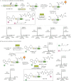
Fig. 6. Incorporation of the 1,2-aminothiol group at any site in proteins for BHoPAL.
a Schematic illustration of our strategy for incorporating the 1,2-aminothiol precursor, thiazolidine, on LAP peptide by employing the LplA enzyme. The structures of LA and TzLA analogues (C2–C7Tz) are shown at the top. b The binding poses of LA (magenta) and C7Tz (orange) in the binding pocket of LplAW37V generated by the Schrödinger software. The black dotted lines indicate hydrogen bonds between C7Tz and the protein residues. The numbers next to these lines denote H–bond lengths in Å. c HPLC analyses of the ligation reactions of LA, C2, C4, C5 and C7Tz with the LAP peptide under conditions “A” and “C” (described in sub-section d below). Our entire HPLC data on all TzLA analogues are provided in Supplementary Fig. 34. d Table summarising the % yields of ligation obtained with different analogues at various time points under different conditions. All the ligation products were characterised by HRMS (Supplementary Table 9). e Top: Scheme for incorporating the 1,2-aminothiol moiety within the MBP-LAP protein followed by BHoPAL. Each of the steps was followed by desalting of the resulting protein conjugate using 10 KDa molecular weight cutoff filters. Bottom: The deconvoluted mass spectra of protein conjugates obtained at each step of the above scheme. The observed masses are shown in black and the corresponding calculated masses are in grey within parenthesis. The XIC and the extended MS traces are provided in Supplementary Fig. 37.
Fig. 7
Fig. 7. Concomitant and Tandem dual labelling of proteins via BHoPAL.
a One-step dual protein modification (Concomitant BHoPAL). Top: Schematic illustration of our strategy for performing dual labelling of proteins. Bottom: The deconvoluted mass spectra of protein conjugates obtained at each step of the above scheme. b Step-wise dual protein modification (Tandem BHoPAL). The deconvoluted mass spectra of protein conjugates obtained at each step are shown next to their structures. The observed masses in all the mass spectra are shown in black and the corresponding calculated masses are in grey within parenthesis. The XIC and the extended MS traces of all the protein conjugates are provided in Supplementary Fig. 42.

References

    1. Hoyt EA, Cal PMSD, Oliveira BL, Bernardes GJL. Contemporary approaches to site-selective protein modification. Nat. Rev. Chem. 2019;3:147–171. doi: 10.1038/s41570-019-0079-1. - DOI
    1. Prescher JA, Bertozzi CR. Chemistry in living systems. Nat. Chem. Biol. 2005;1:13–21. doi: 10.1038/nchembio0605-13. - DOI - PubMed
    1. Tamura T, Hamachi I. Chemistry for covalent modification of endogenous/native proteins: from test tubes to complex biological systems. J. Am. Chem. Soc. 2019;141:2782–2799. doi: 10.1021/jacs.8b11747. - DOI - PubMed
    1. Stephanopoulos N, Francis MB. Choosing an effective protein bioconjugation strategy. Nat. Chem. Biol. 2011;7:876–884. doi: 10.1038/nchembio.720. - DOI - PubMed
    1. Zhang G, Zheng SQ, Liu HP, Chen PR. Illuminating biological processes through site-specific protein labeling. Chem. Soc. Rev. 2015;44:3405–3417. doi: 10.1039/C4CS00393D. - DOI - PubMed