. 2024 Jan 30;15(1):100.
doi: 10.1038/s41419-024-06471-6.

Necroptosis does not drive disease pathogenesis in a mouse infective model of SARS-CoV-2 in vivo

Affiliations

Necroptosis does not drive disease pathogenesis in a mouse infective model of SARS-CoV-2 in vivo

Stefanie M Bader et al. Cell Death Dis. .

Abstract

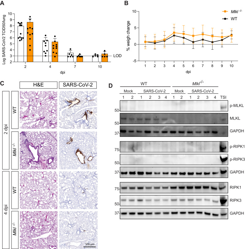
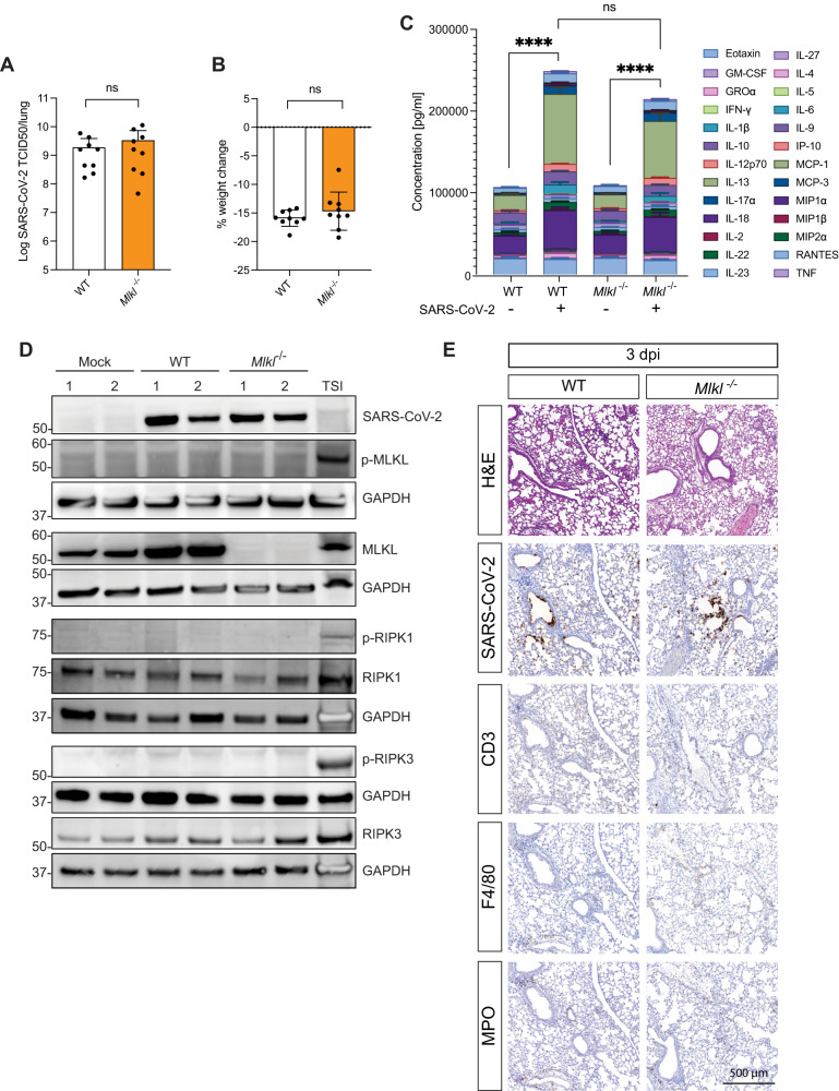
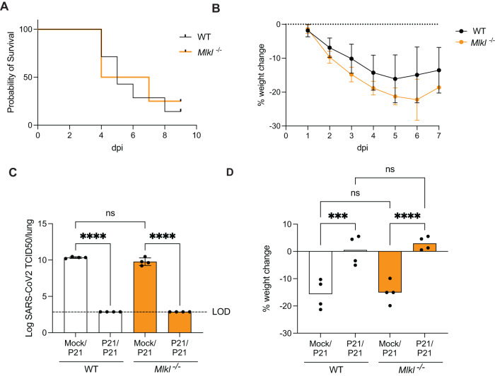
Necroptosis, a type of lytic cell death executed by the pseudokinase Mixed Lineage Kinase Domain-Like (MLKL) has been implicated in the detrimental inflammation caused by SARS-CoV-2 infection. We minimally and extensively passaged a single clinical SARS-CoV-2 isolate to create models of mild and severe disease in mice allowing us to dissect the role of necroptosis in SARS-CoV-2 disease pathogenesis. We infected wild-type and MLKL-deficient mice and found no significant differences in viral loads or lung pathology. In our model of severe COVID-19, MLKL-deficiency did not alter the host response, ameliorate weight loss, diminish systemic pro-inflammatory cytokines levels, or prevent lethality in aged animals. Our in vivo models indicate that necroptosis is dispensable in the pathogenesis of mild and severe COVID-19.


Conflict of interest statement

JMM has received research funding from Anaxis Pharma Pty Ltd. All other authors declare no competing interests.

Figures

Fig. 1
Fig. 1. Necroptosis does not contribute to viral clearance or disease upon infection with a clinical isolate of SARS-CoV-2 (P2).
A, B Mice were challenged intranasally with 104 TCID50 of a clinical SARS-CoV-2 isolate (P2) A Animals were euthanised at defined days post-infection (dpi) and lungs were collected for viral quantification by TCID50 assay. (n = 5–12 mice per group, pooled from 2 independent experiments) B Daily percent weight change of infected animals compared to initial weight. (n = 4–6 mice per group) C Lungs were collected and fixed for histological analysis at 2 and 4 dpi. Representative images of hematoxylin and eosin (H&E) and immunohistochemistry (IHC) stained lungs with SARS-CoV-2 nucleocapsid. Histological images are representative of at least 3 animals. Scale bar = 500 µm. D Western blot analysis of homogenized lungs from wild-type (WT) and MLK knockout (Mlkl−/−) animals 3 days after P2 infection. Samples were probed for phosphorylated MLKL (p-MLKL), MLKL, Glyceraldehyde 3-phosphate dehydrogenase (GAPDH) as a house-keeping gene, phosphorylated RIPK1 (p-RIPK1), phosphorylated RIPK3 (p-RIPK3), RIPK1 and RIPK3. Whole cell lysates of bone marrow derived macrophages treated with Tumor Necrosis Factor (TNF), Smac mimetic IAP antagonist and pan-caspase inhibitor were used as a positive controls for necroptosis (n = 4 mice per group and are representative of 2 independent experiments).
Fig. 2
Fig. 2. Severe disease caused by a mouse-adapted strain of SARS-CoV-2 (P21) is not affected by Mlkl knockout.
A, B, C WT and Mlkl−/− mice were challenged intranasally with 104 TCID50 of SARS-CoV-2 P21 and monitored 3 days post infection for A lung viral burden using TCID50 assay B percent weight change compared to initial weight (n = 9 mice per group), and C the sum of twenty-six cytokines/chemokines expressed in supernatants of lung homogenates (n = 4 mice per group; mean ± SD of each cytokine are shown). D Western blot analysis of homogenized lungs from mock (intranasal DMEM inoculation), wild-type (WT) and MLK knockout (Mlkl−/−) animals 3 days after intranasal P21 infection (10,000 TCID50). Samples were probed for phosphorylated MLKL (p-MLKL), MLKL, Glyceraldehyde 3-phosphate dehydrogenase (GAPDH) as a house-keeping gene, phosphorylated RIPK1 (p-RIPK1), phosphorylated RIPK3 (p-RIPK3), RIPK1, RIPK3 and SARS-CoV-2 nucleocapsid. Whole cell lysates of bone marrow derived macrophages treated with Tumor Necrosis Factor (TNF), Smac mimetic IAP antagonist (LCL-161) and pan-caspase inhibitor (IDN-6556) were used as positive controls for necroptosis (n = 2 mice per group and are representative of 2 independent experiments). E Representative images of hematoxylin and eosin (H&E) and immunohistochemistry (IHC) stained lungs with SARS-CoV-2 nucleocapsid. Mice were challenged intranasally with 104 TCID50 of SARS-CoV-2 P21 and lungs were collected and fixed for histological analysis at 3 dpi. Histological images are representative of at least 3 animals. Scale bars = 500 µm. Unpaired two-tailed student’s t-test (B) after performing log10 transformation (A) and one-way ANOVA with multiple comparisons (C).
Fig. 3
Fig. 3. Lack of necroptosis pathways does not affect age-related disease severity or response to re-challenge.
A, B Aged C57BL/6 and Mlkl−/− mice (6–8 months) were intranasally inoculated with 104 TCID50 SARS-CoV-2 P21 and monitored for A the proportion of mice that became moribund, reaching humane endpoint and B daily percentual weight change over time, relative to the initial weight. (n = 6–8 mice per group; Data is representative of 2 independent experiments) C, B Mock or P21-infected C57BL/6 and Mlkl−/− mice were rechallenged 28 days later with P21 and analyzed 3 days post re-challenge for C lung viral load (TCID50) and D percent weight change, compared to weight before second infection (n = 4 mice per group; Data are representative of 2 independent experiments). One-way ANOVA with multiple comparisons (D), after log10 transformation (C) was performed. Mean ± SD are shown. ***p < 0.001, ****p < 0.0001.

Similar articles

Cited by

References

    1. Singh S, McNab C, Olson RM, Bristol N, Nolan C, Bergstrøm E, et al. How an outbreak became a pandemic: a chronological analysis of crucial junctures and international obligations in the early months of the COVID-19 pandemic. Lancet. 2021;398:2109–24. doi: 10.1016/S0140-6736(21)01897-3. - DOI - PMC - PubMed
    1. Lee S, Channappanavar R, Kanneganti T-D. Coronaviruses: innate immunity, inflammasome activation, inflammatory cell death, and cytokines. Trends Immunol. 2020;41:1083–99. doi: 10.1016/j.it.2020.10.005. - DOI - PMC - PubMed
    1. Morais Da Silva M, Lira De Lucena AS, Paiva Júnior SDSL, Florêncio De Carvalho VM, Santana De Oliveira PS, Rosa MM, et al. Cell death mechanisms involved in cell injury caused by SARS‐CoV‐2. Rev Med Virol. 2021;32:e2292. - PMC - PubMed
    1. Bader SM, Cooney JP, Pellegrini M, Doerflinger M. Programmed cell death: the pathways to severe COVID-19? Biochem J. 2022;479:609–28. doi: 10.1042/BCJ20210602. - DOI - PMC - PubMed
    1. Pasparakis M, Vandenabeele P. Necroptosis and its role in inflammation. Nature. 2015;517:311–20. doi: 10.1038/nature14191. - DOI - PubMed

Publication types

Substances