. 2024 Jan 29;15(1):875.
doi: 10.1038/s41467-024-45009-4.

Expanded palette of RNA base editors for comprehensive RBP-RNA interactome studies

Affiliations

Expanded palette of RNA base editors for comprehensive RBP-RNA interactome studies

Hugo C Medina-Munoz et al. Nat Commun. .

Abstract

RNA binding proteins (RBPs) are key regulators of RNA processing and cellular function. Technologies to discover RNA targets of RBPs such as TRIBE (targets of RNA binding proteins identified by editing) and STAMP (surveying targets by APOBEC1 mediated profiling) utilize fusions of RNA base-editors (rBEs) to RBPs to circumvent the limitations of immunoprecipitation (CLIP)-based methods that require enzymatic digestion and large amounts of input material. To broaden the repertoire of rBEs suitable for editing-based RBP-RNA interaction studies, we have devised experimental and computational assays in a framework called PRINTER (protein-RNA interaction-based triaging of enzymes that edit RNA) to assess over thirty A-to-I and C-to-U rBEs, allowing us to identify rBEs that expand the characterization of binding patterns for both sequence-specific and broad-binding RBPs. We also propose specific rBEs suitable for dual-RBP applications. We show that the choice between single or multiple rBEs to fuse with a given RBP or pair of RBPs hinges on the editing biases of the rBEs and the binding preferences of the RBPs themselves. We believe our study streamlines and enhances the selection of rBEs for the next generation of RBP-RNA target discovery.


Conflict of interest statement

G.W.Y. is an SAB member of Jumpcode Genomics and a co-founder, member of the Board of Directors, on the SAB, equity holder, and paid consultant for Locanabio (until 31 December 2023) and Eclipse BioInnovations. G.W.Y. is a distinguished visiting professor at the National University of Singapore. G.W.Y.’s interests have been reviewed and approved by the University of California San Diego in accordance with its conflict-of-interest policies. A.C.K. is a member of the SAB of Pairwise Plants, is an equity holder for Pairwise Plants and Beam Therapeutics, and receives royalties from Pairwise Plants, Beam Therapeutics, and Editas Medicine via patents licensed from Harvard University. A.C.K.’s interests have been reviewed and approved by the University of California San Diego in accordance with its conflict-of-interest policies. Other authors declare no competing interests.

Figures
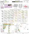
Fig. 1
Fig. 1. A reporter system to assay RNA base editing activity of candidate RNA base editors (rBEs).
a Reporter mRNA bearing twelve MS2 bacteriophage stem-loops (yellow bars) downstream of a Super folder GFP coding sequence (sfGFP) and a chimera bearing an MS2 coat-protein (MCP, black) domain fused to a candidate rBE (brown). b Strategy to test candidate rBEs. Plasmids encoding the constructs in a) are co-transfected into HEK293XT cells so that the MCP binds the MS2 stem-loops in the reporter, and the rBE catalyzes RNA editing. After total RNA is isolated, targeted RNA sequencing is used to detect edits along the reporter sequence. c Fraction of total covered bases at each position along the twelve MS2 stem-loop reporters exhibiting either C-to-U (green), A-to-I (orange), or no (black horizontal line) edits. d The number of C-to-U (green) and A-to-I (orange) edits on the reporter mRNA (on-target, left) or other poly(A)+ RNAs (off-target, right). e Ratio of the number of on-target C-to-U (green) and A-to-I (orange) edits on twelve MS2 loop construct vs. the number of off-target edits on poly(A)+ RNAs, plotted for n = 2 independent experiments across each enzyme. Boxes extend from the first to the third quartile of the data, with the center line indicating the median. Box whiskers extend to the farthest data point lying within 1.5x the inter-quartile range from the box in either direction. (Figure created with BioRender).
Fig. 2
Fig. 2. RBP-Mediated RNA Editing with RBFOX2 and Top rBE Candidates.
a Constructs feature RBFOX2 RNA-binding protein (RBP, pink) fused to RNA base editors (rBEs, brown) at the C-terminal end (“Sequence-specific RBP-associated rBEs”) and solo rBEs (“Free editors”). b RBFOX2-directed or free rBE activity detection in HEK293XT cells involves plasmid transfection. RBFOX2-RBP fused rBEs target GCAUG binding sites, differing from free rBEs. After 72 h, RNA is extracted for poly(A) + RNA sequencing and FLARE edit cluster detection. c, d FLARE analysis reveals edit clusters and RNA species edited by each RBFOX2-rBE fusion. Colors: RBFOX2 fusion to 8e (dark blue), A2dd (R) (teal), APOBEC1 (green), 7.10 (V82G) (red). e HOMER analysis identifies the canonical RBFOX2 binding-site motif ((U)GCAUG) as the top motif in each fusion’s edit clusters, using a cumulative hypergeometric distribution for p-values. f Replicable edit cluster fraction in RBFOX2-rBE fusions (n = 3 experiments) is higher than in random edit clusters from mRNA targets, showing enrichment. Box plots display data, with boxes from first to third quartiles, median center line, and whiskers extending to 1.5x the inter-quartile range. g Density plots show RBFOX2-rBE fusions’ replicable peak centers (n = 3 experiments) closer to the RBFOX2 motif than rBEs alone. RBFOX2-rBE fusion colors as in 2c; Free rBE colors distinct: 8e (purple), A2dd (R) (turquoise), APOBEC1 (light green), 7.10 (V82G) (pink). h Fraction of replicable edit clusters in RBFOX2-rBE fusions overlapping RBFOX2-APOBEC1 eCLIP peaks is higher than in random peak sets. Box plot details as in (f). i, j Analysis of relationships between edit cluster sets and RNA species for each RBFOX2-rBE fusion is depicted with grey dots. Lines bisect intersecting values, and black bars reflect set numbers. k The canonical RBFOX2 binding-site motif is predominant in unique clusters from RBFOX2-rBE fusion, as per HOMER analysis. (Figure created with BioRender).
Fig. 3
Fig. 3. Analysis of RBFOX2-rBE and free rBE sequence biases.
a A CNN with two convolutional layers identifies enzyme-specific biases in enzyme-RBFOX2 fusion and enzyme-alone experiments. Higher AUCs for enzymes alone suggest more identifiable biases. Color-coding: RBFOX2 fusions with 8e (dark blue), A2dd (R) (teal), APOBEC1 (green), and 7.10 (V82G) (red). For enzymes alone: 8e (purple), A2dd (R) (turquoise), APOBEC1 (light green), and 7.10 (V82G) (pink). b A CNN trained on enzyme-alone clusters predicts enzyme identity in fusion peaks well, often outperforming one trained on fusion clusters. Conversely, a CNN trained on enzyme-fusion clusters is less effective on free RBE clusters (right). c Edited sites show distinct, replicable enzyme-specific flanking base context preferences, evident in a PCA plot, whether alone or fused to RBFOX2. d In the PCA plot, the first (black) and second (purple) principal components distinctly reflect contributions from each RNA base or combinations (e.g., GC). e Density plots reveal consistent peak adenosine (A, left) and guanosine-cytidine (GC) content across RBFOX2-rBE and free rBE (top and bottom, respectively) for any enzyme. f Edit site context preferences, indicated by the height of each bar, vary by enzyme and are consistent between free and RBFOX2-rBE edits. Each flanking base set is color-coded to match its bar in the chart. g On genes with two GCAUG motifs edited by different enzymes, peak base contents and enzyme editing context specificities (from e) influence editing likelihood for each region. h Comparison of combined edit clusters (red bar) and individual RBFOX2-rBE edit cluster sets intersecting with RBFOX2-APOBEC1 eCLIP peak sequences. i Boxplots compare RBFOX2-APOBEC1 eCLIP peak sequence overlaps with replicable RBFOX2-rBE edit clusters against thirty random eCLIP sets; red circles for actual peaks, enrichment shown as red bars. Boxes cover middle 50% of data, median as center line, whiskers up to 1.5x inter-quartile range.
Fig. 4
Fig. 4. RNA Editing as a Proxy for Translation with RPS2 and Top rBE Candidates.
a Construct with small ribosomal subunit protein RPS2 (purple) fused to a candidate rBE (brown) at its C-terminal end (“Translation-directed editor”). b RPS2-directed editing detection involves transfecting HEK293XT cells with a plasmid encoding the above construct and incubating with Torin-1 to inhibit translation or DMSO as a control. Post-treatment, poly(A) + RNA libraries are prepared and sequenced. Torin-1 is predicted to reduce editing, as indicated by reduction in edits per read (EPR). c EPR in Torin-1 treated cells shows a reduction compared to DMSO-treated cells across all RPS2-rBE fusions. d Statistically significant changes in RPS2-rBE EPR post Torin-1 treatment: decrease (red), increase (blue), or no significant change (grey) compared to DMSO control. e Log2-transformed decrease in EPR due to Torin-1, calculated as EPR in Torin-1 condition over untreated, across n = 3 experiments for RPS2-enzyme fusions in poly(A)+ RNAs (black) and TOP-containing mRNAs (pink). Boxes span the middle 50% of the data. The median is represented as the center line, and whiskers extend to the farthest data point lying within 1.5x the inter-quartile range from the box in either direction. f Edits per read fold-change between Torin-1- and DMSO-treated cells and associated statistical significance (log2-transformed p-value) for each RNA. Poly(A)+ RNAs (black) and TOP-containing mRNAs (pink) are highlighted. A two-sided t-test was used to determine significance. g Boxplots reveal EPR differences between Torin-1 and DMSO treatments in coding sequences (CDS, purple shading) and 3’ UTRs, over n = 3 experiments. Pronounced changes in CDS-specific editing for poly(A)+ RNAs (black) and TOP mRNAs (pink). Boxes cover middle 50%, median as center line, whiskers up to 1.5x inter-quartile range. (Figure created with BioRender).
Fig. 5
Fig. 5. A combinatorial editing reporter system to identify multiple RBP associations with the same transcript.
a Components that are used to test combinatorial C-to-U and A-to-I rBE pairs. The system uses a reporter mRNA with varied MS2- (yellow rectangle) and PP7- (red rectangle) stem-loop distributions in the 3’ UTR. The reporter binds the MS2 (MCP, black) and PP7 (red) coat proteins fused to C-to-U and A-to-I rBEs. b A combinatorial editing strategy. Plasmids encoding C-to-U and A-to-I fused MCP and PP7 proteins are co-transfected into HEK293XT cells with the reporter bearing both binding sites. After, the edits are detected on the reporter with targeted RNA sequencing. ce Distribution of C-to-U (green) and A-to-I (orange) edits deposited by each of five different enzyme combinations and the reporter without enzymes. The edits are mapped along each of the three distinct reporters. One reporter bears a c MS2 (yellow) and PP7 (red) stem-loops spaced 50 bp apart, with 350 bp separating the pairs. The other two reporters contain d twelve or e four alternating MS2 and PP7 binding sites spaced 50 bp apart. (Figure created with BioRender).

Update of

References

    1. Gebauer F, Schwarzl T, Valcárcel J, Hentze MW. RNA-binding proteins in human genetic disease. Nat. Rev. Genet. 2021;22:185–198. doi: 10.1038/s41576-020-00302-y. - DOI - PubMed
    1. Ingolia NT, Ghaemmaghami S, Newman JRS, Weissman JS. Genome-wide analysis in vivo of translation with nucleotide resolution using ribosome profiling. Science. 2009;324:218–223. doi: 10.1126/science.1168978. - DOI - PMC - PubMed
    1. Van Nostrand EL, et al. Robust transcriptome-wide discovery of RNA-binding protein binding sites with enhanced CLIP (eCLIP) Nat. Methods. 2016;13:508–514. doi: 10.1038/nmeth.3810. - DOI - PMC - PubMed
    1. McMahon AoifeC, et al. TRIBE: hijacking an RNA-editing enzyme to identify cell-specific targets of RNA-binding proteins. Cell. 2016;165:742–753. doi: 10.1016/j.cell.2016.03.007. - DOI - PMC - PubMed
    1. Brannan KW, et al. Robust single-cell discovery of RNA targets of RNA-binding proteins and ribosomes. Nat. Methods. 2021;18:507–519. doi: 10.1038/s41592-021-01128-0. - DOI - PMC - PubMed

MeSH terms