. 2024 Feb;30(2):394-402.
doi: 10.1038/s41591-023-02729-2. Epub 2024 Jan 29.

Iatrogenic Alzheimer's disease in recipients of cadaveric pituitary-derived growth hormone

Affiliations

Iatrogenic Alzheimer's disease in recipients of cadaveric pituitary-derived growth hormone

Gargi Banerjee et al. Nat Med. 2024 Feb.

Abstract

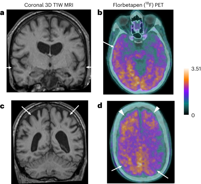
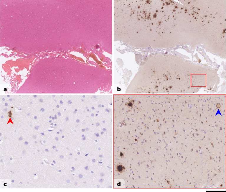
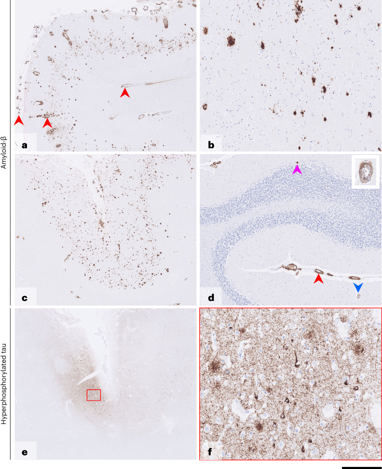
Alzheimer's disease (AD) is characterized pathologically by amyloid-beta (Aβ) deposition in brain parenchyma and blood vessels (as cerebral amyloid angiopathy (CAA)) and by neurofibrillary tangles of hyperphosphorylated tau. Compelling genetic and biomarker evidence supports Aβ as the root cause of AD. We previously reported human transmission of Aβ pathology and CAA in relatively young adults who had died of iatrogenic Creutzfeldt-Jakob disease (iCJD) after childhood treatment with cadaver-derived pituitary growth hormone (c-hGH) contaminated with both CJD prions and Aβ seeds. This raised the possibility that c-hGH recipients who did not die from iCJD may eventually develop AD. Here we describe recipients who developed dementia and biomarker changes within the phenotypic spectrum of AD, suggesting that AD, like CJD, has environmentally acquired (iatrogenic) forms as well as late-onset sporadic and early-onset inherited forms. Although iatrogenic AD may be rare, and there is no suggestion that Aβ can be transmitted between individuals in activities of daily life, its recognition emphasizes the need to review measures to prevent accidental transmissions via other medical and surgical procedures. As propagating Aβ assemblies may exhibit structural diversity akin to conventional prions, it is possible that therapeutic strategies targeting disease-related assemblies may lead to selection of minor components and development of resistance.


Conflict of interest statement

J.C. is a shareholder and director of D-Gen, Ltd., an academic spin-out company working in the field of prion disease diagnosis, decontamination and therapeutics. D-Gen supplied the ICSM35 antibody used for PrP immunohistochemistry. The other authors declare no competing interests.

Figures

Fig. 1
Fig. 1. Magnetic resonance and amyloid-PET (18F-Florbetapen) images—case 3.
a, High-resolution three-dimensional (3D) T1-weighted (T1W) magnetic resonance (MR) coronal image through the temporal lobes demonstrates volume loss within the temporal lobes bilaterally (arrows) and also marked central atrophy. b, Axial PET images demonstrate diffuse increased tracer uptake in the cortex and subcortical white matter, increased in the right temporal lobe compared to the left. c, High-resolution MR (3D T1W) coronal image through superior parietal lobules bilaterally demonstrates marked volume loss (arrows). d, Axial PET images demonstrate marked tracer uptake within the superior parietal lobules bilaterally (arrows) in addition to increased uptake in the bilateral frontal lobes (arrowheads).
Fig. 2
Fig. 2. Brain biopsy—case 2.
Images shown are from a left frontal lobe brain biopsy. H&E-stained preparation (a) shows full-thickness well-preserved cortical hexa-laminar cytoarchitecture with unremarkable overlying leptomeninges. Immunostaining for Aβ (b and d) shows frequent diffuse parenchymal deposits with no plaques with central amyloid cores and a single blood vessel with concentric Aβ angiopathy but no associated inflammation. Hyperphosphorylated tau (c) is restricted to rare dystrophic deposits, with no evidence of neuronal or glial tau pathology. Brain postmortem findings are provided in the Supplementary Information. Scale bar, 750 µm in a and b, 50 µm in c and 100 µm in d. Aβ antibody: clone 6F3D, dilution 1:50, source DAKO, product number M0872. Hyperphosphorylated tau antibody: clone AT8, dilution 1:1,200, source Invitrogen (Thermo Fisher Scientific), product number MN1020.
Fig. 3
Fig. 3. Postmortem brain tissue—case 1.
Immunostaining for Aβ (ad) shows frequent parenchymal deposits in the cortex (a and c) and caudate nucleus (b), with rare, isolated deposits in the cerebellar cortex (d, pink arrowhead). In the cerebrum (a and c), there is widespread, concentric amyloid angiopathy in the leptomeninges, cortex and subcortical white matter (red arrowheads in a), and, in the cerebellum (d), there is widespread concentric amyloid angiopathy in the leptomeninges (red arrowhead) and occasionally in the cerebellar cortex (blue arrowhead; inset shows vessel at higher magnification), without associated inflammation. Immunostaining for hyperphosphorylated tau (AT8) of the insular cortex (e and f) shows pan-cortical patches of a dense meshwork of neuropil threads, frequent pre-tangles, occasional tangles and moderately frequent neuritic plaques. Scale bar, 1.5 mm in a, 250 µm in b, 170 µm in c, 400 µm in d, 1.8 mm in e and 130 µm in f. Aβ antibody: clone 6F3D, dilution 1:50, source DAKO, product number M0872. Hyperphosphorylated tau antibody: clone AT8, dilution 1:1,200, source Invitrogen (Thermo Fisher Scientific), product number MN1020.
Extended Data Fig. 1
Extended Data Fig. 1. Post-mortem brain tissue, Case 2.
Haematoxylin and eosin-stained preparation (a) demonstrates extensively calcified and ossified adamantinomatous craniopharyngioma invading into the brain tissue, without any histological signs of malignant transformation. Immunostaining for amyloid-β shows no evidence of pathology presence in the hippocampal region and parahippocampal gyrus (b) or basal ganglia, brainstem and cerebellum (not shown). Hyperphosphorylated tau pathology in the limbic region is restricted to rare isolated neurofibrillary tangles, pre-tangles and threads in the entorhinal cortex (c and d, with blue arrowheads in D). Scale bar: 230µm in A; 3mm in B and c, and 220µm in d. Amyloid-β antibody: clone 6F3D, dilution 1:50, source DAKO, product number M0872. Hyperphosphorylated tau antibody: clone AT8, dilution 1:1200, source Invitrogen (Thermo), product number MN1020.

References

    1. Collinge J. Mammalian prions and their wider relevance in neurodegenerative diseases. Nature. 2016;539:217–226. - PubMed
    1. Manka SW, et al. A structural basis for prion strain diversity. Nat. Chem. Biol. 2023;19:607–613. - PMC - PubMed
    1. Collinge J, et al. Kuru in the 21st century—an acquired human prion disease with very long incubation periods. Lancet. 2006;367:2068–2074. - PubMed
    1. Mead S, et al. Balancing selection at the prion protein gene consistent with prehistoric kurulike epidemics. Science. 2003;300:640–643. - PubMed
    1. Collinge J, Sidle KC, Meads J, Ironside J, Hill AF. Molecular analysis of prion strain variation and the aetiology of ‘new variant’ CJD. Nature. 1996;383:685–690. - PubMed