. 2024 Jan 29;25(1):36.
doi: 10.1186/s13059-024-03173-2.

Mosaic loss of Y chromosome is associated with aging and epithelial injury in chronic kidney disease

Affiliations

Mosaic loss of Y chromosome is associated with aging and epithelial injury in chronic kidney disease

Parker C Wilson et al. Genome Biol. .

Abstract

Background: Mosaic loss of Y chromosome (LOY) is the most common chromosomal alteration in aging men. Here, we use single-cell RNA and ATAC sequencing to show that LOY is present in the kidney and increases with age and chronic kidney disease.

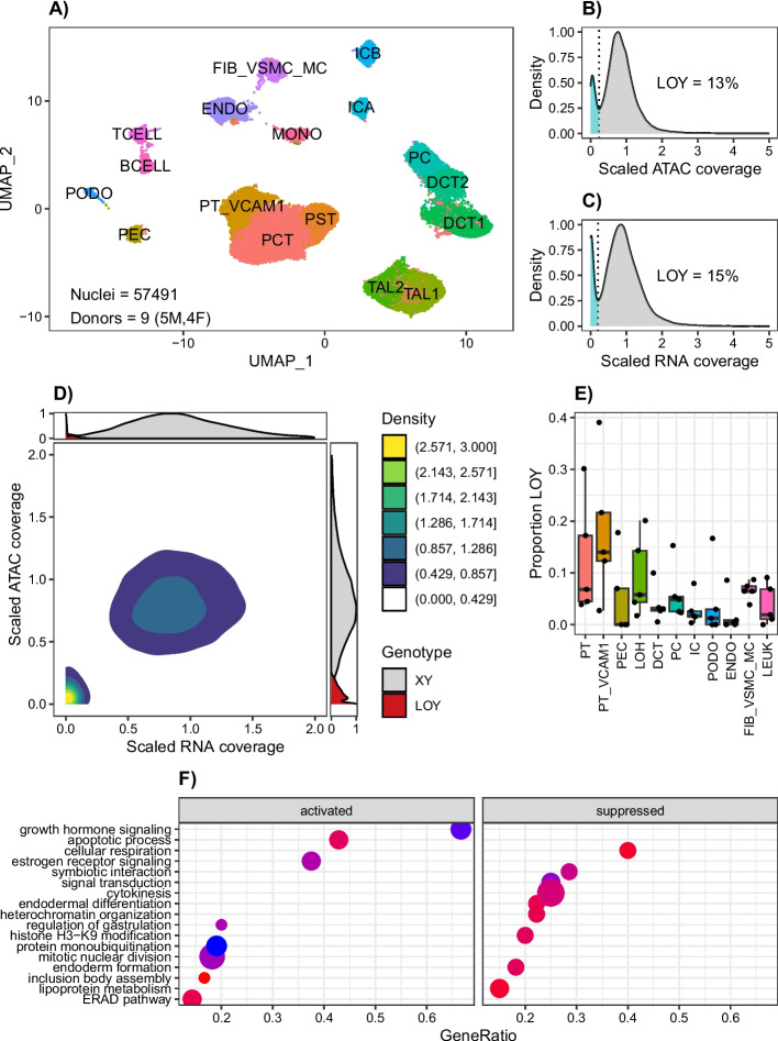
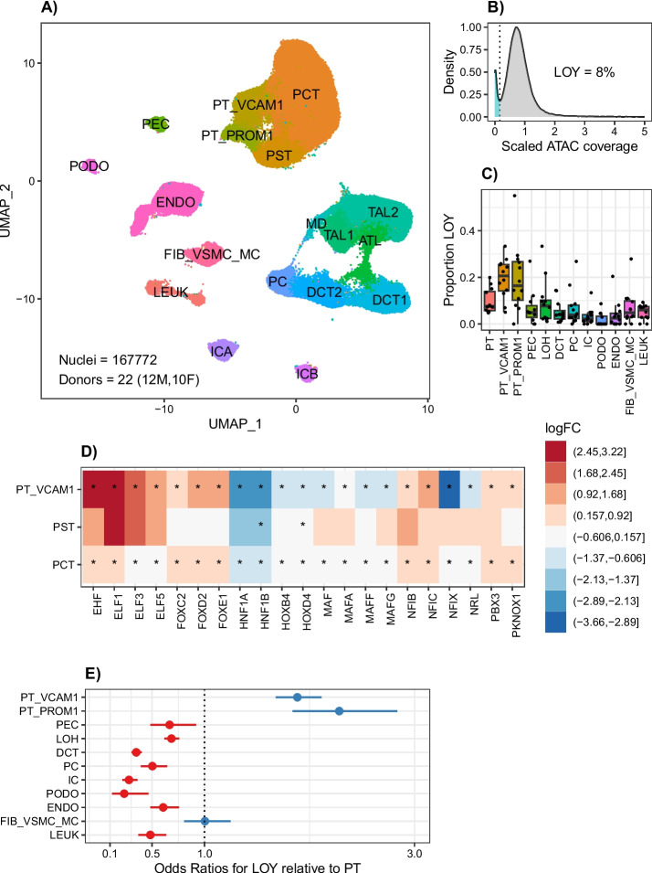
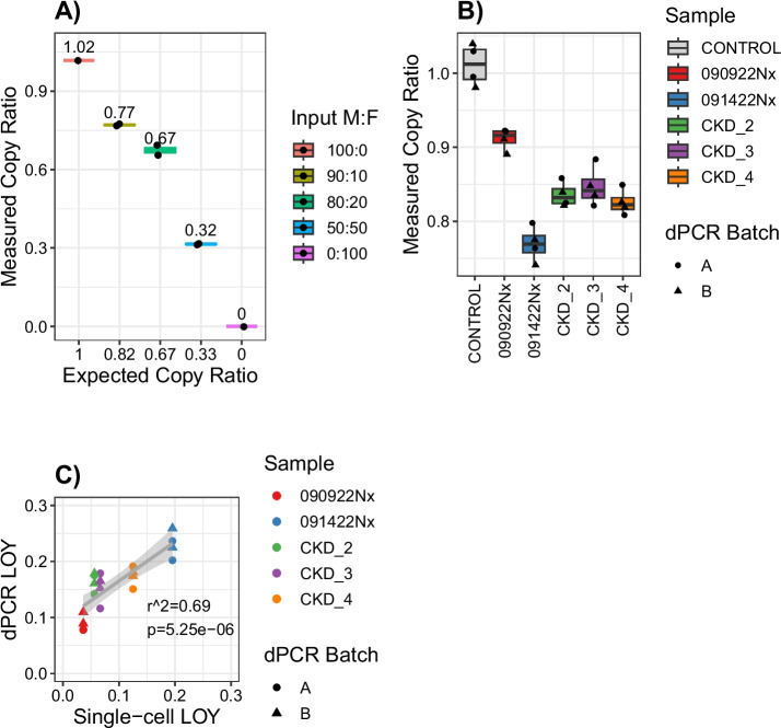
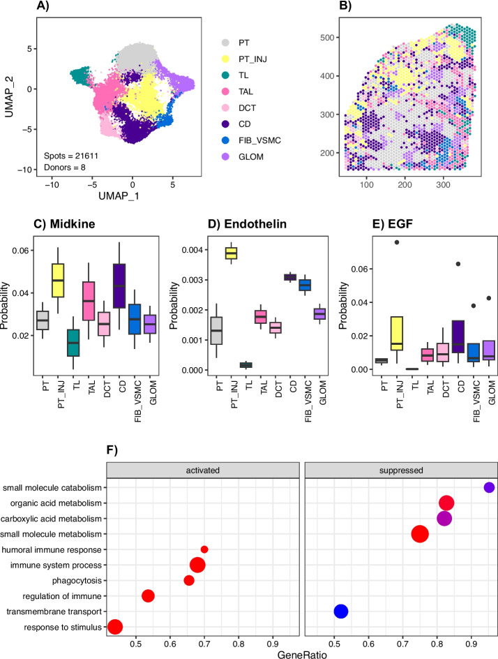
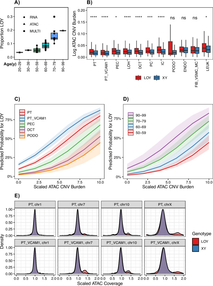
Results: The likelihood of a cell having LOY varies depending on its location in the nephron. Cortical epithelial cell types have a greater proportion of LOY than medullary or glomerular cell types, which may reflect their proliferative history. Proximal tubule cells are the most abundant cell type in the cortex and are susceptible to hypoxic injury. A subset of these cells acquires a pro-inflammatory transcription and chromatin accessibility profile associated with expression of HAVCR1, VCAM1, and PROM1. These injured epithelial cells have the greatest proportion of LOY and their presence predicts future kidney function decline. Moreover, proximal tubule cells with LOY are more likely to harbor additional large chromosomal gains and express pro-survival pathways. Spatial transcriptomics localizes injured proximal tubule cells to a pro-fibrotic microenvironment where they adopt a secretory phenotype and likely communicate with infiltrating immune cells.

Conclusions: We hypothesize that LOY is an indicator of increased DNA damage and potential marker of cellular senescence that can be applied to single-cell datasets in other tissues.


Conflict of interest statement

B.D.H. is a consultant for Janssen Research & Development, LLC, Pfizer, and Chinook Therapeutics and holds equity in Chinook Therapeutics and grant funding from Janssen Research & Development, LLC, and Pfizer; all interests are unrelated to the current work.

Figures

Fig. 1
Fig. 1
LOY detection by single nucleus multiome sequencing. A UMAP of male and female kidney cell types. PCT, proximal convoluted tubule; PST, proximal straight tubule; PT_VCAM1, VCAM1 + proximal tubule; PEC, parietal epithelial cells; TAL1, cortical thick ascending limb; TAL2, medullary thick ascending limb; DCT1, early distal convoluted tubule; DCT2, late distal convoluted tubule; PC, principal cells; ICA, type A intercalated cells; ICB, type B intercalated cells; PODO, podocytes; ENDO, endothelial cells; FIB_VSMC_MC, fibroblasts; vascular smooth muscle; and mesangial cells; TCELL, T cells; BCELL, B cells; MONO, mononuclear cells. B ATAC modality LOY density plot in 23,333 male cells. C RNA modality LOY density plot in 23,333 male cells D Joint ATAC/RNA LOY density plot in 23,333 male cells. E Median proportion LOY in male kidney cell types. F Gene ontology pathway enrichment for age-adjusted differentially expressed genes associated with LOY in the male proximal tubule
Fig. 2
Fig. 2
snATAC-seq detection of LOY. A UMAP of male and female kidney cell types PCT, proximal convoluted tubule; PST, proximal straight tubule; PT_VCAM1, VCAM1 + proximal tubule; PT_PROM1, PROM1 + proximal tubule; PEC, parietal epithelial cells; ATL, ascending thin limb; TAL1, cortical thick ascending limb; TAL2, medullary thick ascending limb; MD, macula densa; DCT1, early distal convoluted tubule; DCT2, late distal convoluted tubule; PC, principal cells; ICA, type A intercalated cells; ICB, type B intercalated cells; PODO, podocytes; ENDO, endothelial cells; FIB_VSMC_MC, fibroblasts, vascular smooth muscle, and mesangial cells; LEUKleukocytes. B ATAC modality LOY density plot in 47,458 male cells. C Median proportion LOY in male kidney cell types. D Log-fold-change transcription factor activity for LOY vs. XY in male kidney cell types for transcription factors with differential activity in at least two proximal tubule subpopulations. F GLMM for LOY in male kidney cell types
Fig. 3
Fig. 3
LOY detection by scRNA-seq. A UMAP of male and female kidney cell types PT, proximal tubule; PT_VCAM1, VCAM1 + proximal tubule; PT_PROM1, PROM1 + proximal tubule; PT_MT, mitochondrial gene proximal tubule; PEC, parietal epithelial cells; LOH, loop of Henle; DCT, distal convoluted tubule; PC, principal cells; IC, intercalated cells; PODO, podocytes; ENDO, endothelial cells; FIB_VSMC_MC, fibroblasts; vascular smooth muscle, and mesangial cells, LEUKleukocytes. B RNA modality LOY density plot in 78,738 male cells. C Median proportion LOY in male kidney cell types. D Log-fold-change gene expression for LOY vs. XY in male kidney cell types. E Proportion LOY in Control or AKI vs CKD male samples. F GLMM for LOY in male kidney cell types adjusted for CKD
Fig. 4
Fig. 4
LOY detection by digital PCR. A gDNA was extracted from male hTERT-RPTEC or female HEK293T cell lines and mixed in the indicated male: female (M:F) ratios. The mixed DNA samples were measured by multiplex dPCR using a FAM-labeled chrY target and ABY-labeled chrX reference. The measured copy ratio is the number of microwells that amplify the chrY target divided by the number of microwells that amplify the chrX reference. These values were compared to the expected copy ratio, which was estimated based on the relative ratio of M:F DNA input. For example, a 50:50 mixture of M:F DNA would yield 1 copy of chrY for every 3 copies of chrX and an expected copy ratio of 0.33. B gDNA was extracted from male kidney cortex samples and assayed by dPCR as previously described. Each sample was assayed with 2 technical replicates and 2 independent batches for a total of 4 measurements per sample. The sample labeled control is gDNA isolated from a male hTERT-RPTEC cell line. C We used dPCR to estimate the fraction of male cells with LOY using the following formula: 1 − chrY/chrX. This approach assumes the concentration of chrX is constant and that any changes in the measured copy ratio are due to changes in the concentration of chrY. We compared our dPCR and single-cell estimates to compute an r-squared value using the lm function in R
Fig. 5
Fig. 5
Spatial transcriptomics in the kidney identifies a pro-fibrotic microenvironment. A UMAP of integrated spatial datasets with neighborhood annotations PT, proximal tubule; PT_INJ, injured proximal tubule; TL, thin limb; TAL, thick ascending limb; DCT, distal convoluted tubule; CD, collecting duct; FIB_VSMC, fibroblasts and vascular smooth muscle cells; GLOM, glomeruli. B Representative spot annotation and visualization of spatial neighborhoods. C CellChat midkine secreted signaling arising from the PT_INJ neighborhood. D CellChat endothelin secreted signaling arising from the PT_INJ neighborhood. E CellChat EGF secreted signaling arising from the PT_INJ neighborhood. F Gene ontology enrichment for PT_INJ spot-specific differentially expressed genes
Fig. 6
Fig. 6
Age and mCA as Predictors for LOY. A Proportion LOY for single-cell multiomes, snATAC-seq and scRNA-seq for male donors (n = 32) binned by age. B Genome-wide CNV burden for LOY vs XY in male single-cell multiomes and snATAC-seq datasets. C Marginal probabilities for LOY by cell type and CNV burden in male single-cell multiomes and snATAC-seq. D Marginal probabilities for LOY by age and CNV burden in male single-cell multiomes and snATAC-seq. E Coverage density for male kidney cell types by chromosome in LOY and XY cells

References

    1. Forsberg LA, Gisselsson D, Dumanski JP. Mosaicism in health and disease — clones picking up speed. Nat Rev Genet. 2017;18(2):128–142. doi: 10.1038/nrg.2016.145. - DOI - PubMed
    1. Solís-Moruno M, Batlle-Masó L, Bonet N, Aróstegui JI, Casals F. Somatic genetic variation in healthy tissue and non-cancer diseases. Eur J Hum Genet. 2023;31(1):48–54. doi: 10.1038/s41431-022-01213-8. - DOI - PMC - PubMed
    1. Liu X, Kamatani Y, Terao C. Genetics of autosomal mosaic chromosomal alteration (mCA) J Hum Genet. 2021;66(9):879–885. doi: 10.1038/s10038-021-00964-4. - DOI - PubMed
    1. Forsberg LA. Loss of chromosome Y (LOY) in blood cells is associated with increased risk for disease and mortality in aging men. Hum Genet. 2017;136(5):657–663. doi: 10.1007/s00439-017-1799-2. - DOI - PMC - PubMed
    1. Jacobs PA, Brunton M, Court Brown WM, Doll R, Goldstein H. Change of human chromosome count distribution with age: evidence for a sex differences. Nature. 1963;197:1080–1081. doi: 10.1038/1971080a0. - DOI - PubMed

Publication types