

Automated localization of mandibular landmarks in the construction of mandibular median sagittal plane
- PMID: 38287445
- PMCID: PMC10823719
- DOI: 10.1186/s40001-024-01681-2
Automated localization of mandibular landmarks in the construction of mandibular median sagittal plane
Abstract
Objective: To use deep learning to segment the mandible and identify three-dimensional (3D) anatomical landmarks from cone-beam computed tomography (CBCT) images, the planes constructed from the mandibular midline landmarks were compared and analyzed to find the best mandibular midsagittal plane (MMSP).
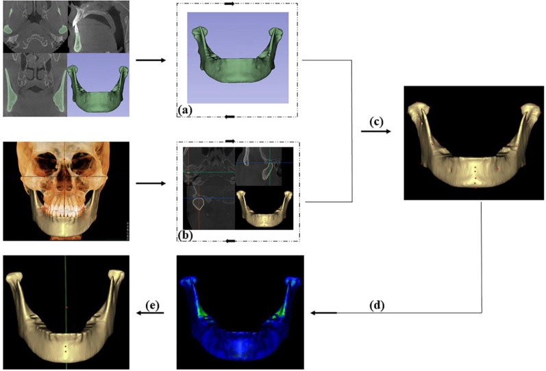
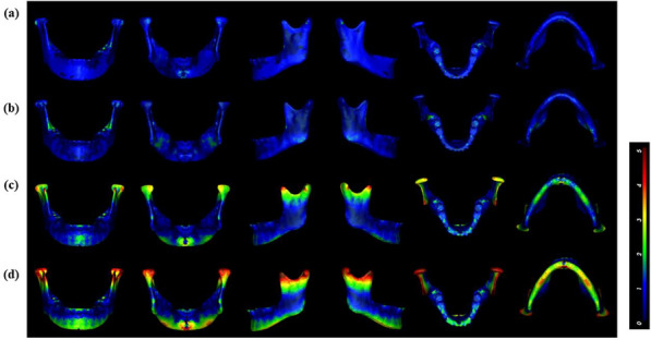
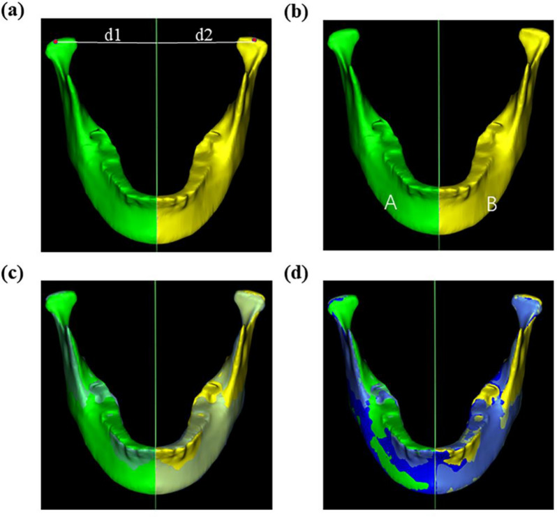
Methods: A total of 400 participants were randomly divided into a training group (n = 360) and a validation group (n = 40). Normal individuals were used as the test group (n = 50). The PointRend deep learning mechanism segmented the mandible from CBCT images and accurately identified 27 anatomic landmarks via PoseNet. 3D coordinates of 5 central landmarks and 2 pairs of side landmarks were obtained for the test group. Every 35 combinations of 3 midline landmarks were screened using the template mapping technique. The asymmetry index (AI) was calculated for each of the 35 mirror planes. The template mapping technique plane was used as the reference plane; the top four planes with the smallest AIs were compared through distance, volume difference, and similarity index to find the plane with the fewest errors.
Results: The mandible was segmented automatically in 10 ± 1.5 s with a 0.98 Dice similarity coefficient. The mean landmark localization error for the 27 landmarks was 1.04 ± 0.28 mm. MMSP should use the plane made by B (supramentale), Gn (gnathion), and F (mandibular foramen). The average AI grade was 1.6 (min-max: 0.59-3.61). There was no significant difference in distance or volume (P > 0.05); however, the similarity index was significantly different (P < 0.01).
Conclusion: Deep learning can automatically segment the mandible, identify anatomic landmarks, and address medicinal demands in people without mandibular deformities. The most accurate MMSP was the B-Gn-F plane.
Keywords: 3D imaging; Deep learning; Mandible segmentation; Mandibular median sagittal plane.
© 2024. The Author(s).
Conflict of interest statement
The authors declare no competing interests.
Figures

References
Publication types
MeSH terms
Grants and funding
LinkOut - more resources
Full Text Sources
