

The pterygomandibular raphe: a comprehensive review
- PMID: 38287643
- PMCID: PMC10968190
- DOI: 10.5115/acb.23.232
The pterygomandibular raphe: a comprehensive review
Abstract
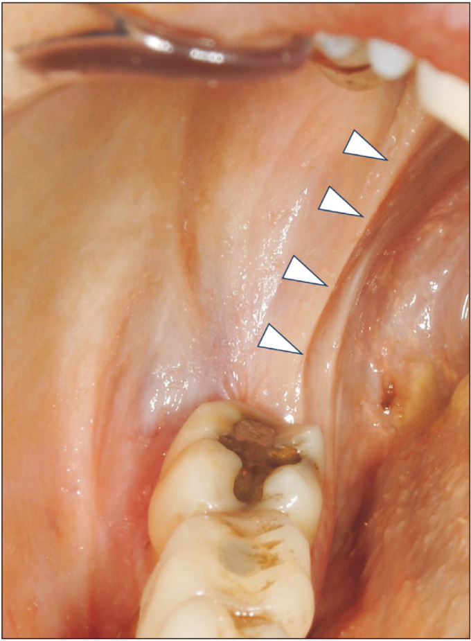
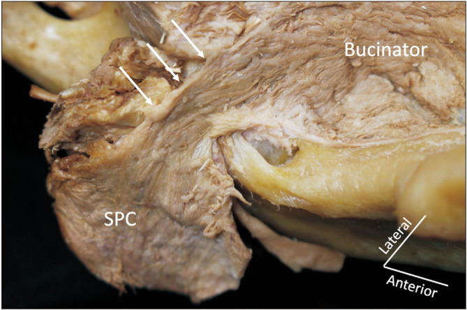
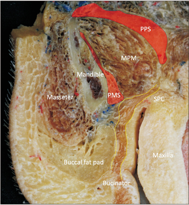
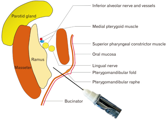
The pterygomandibular raphe (PMR) is a tendinous structure connecting the bucinator and the superior pharyngeal constrictor muscles. With its implications in the spread of oral cancer, the proper treatment of obstructive sleep apnea, and dental procedures, it is important to obtain a thorough understanding of the PMR. We reviewed the existing literature to compile the published information regarding its anatomy, embryology, imaging, variations, functions, pathologies, and clinical relevance of the pterygomandibular raphe.
Keywords: Anatomic variation; Anatomy; Embryology; Oral; Tendons.
Conflict of interest statement
No potential conflict of interest relevant to this article was reported.
Figures
References
-
- Macalister A. A text-book of human anatomy: systematic and topographical, including the embryology, histology and morphology of man, with special reference to the requirements of practical surgery and medicine. Griffin; 1889.
-
- Hollstein L. In: [Textbook of human anatomy] Verlag von E.H, editor. Schroeder; 1873. p. 279. German.
-
- Poirier P, Charpy A. [Treatise on human anatomy] 2nd-3rd ed. Masson; 1901. pp. 355–7. French.
Publication types
LinkOut - more resources
Full Text Sources