

A late presentation of a left paraduodenal hernia in an elderly patient admitted in emergency: A case report
- PMID: 38288049
- PMCID: PMC10823032
- DOI: 10.1016/j.radcr.2024.01.013
A late presentation of a left paraduodenal hernia in an elderly patient admitted in emergency: A case report
Abstract
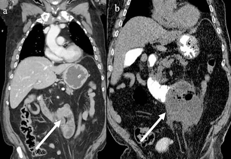
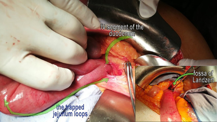
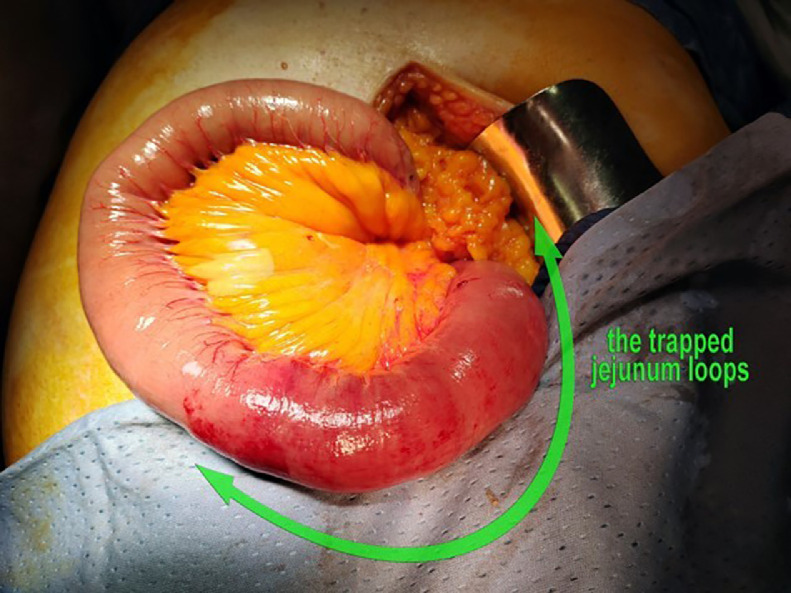
Small bowel internal hernias (IHs), a rare cause of small bowel occlusion (SBO) and small bowel strangulation, while more commonly seen in young adults, can also affect elderly patients and pose a significant diagnostic challenge due to their nonspecific symptoms. In most cases, laparotomy was used to diagnose IHs. However, multidetector computed tomography (MDCT) is usually the best imaging tool to use in the emergency setting for the diagnosis of IHs. An 83-year-old man was admitted to emergency with acute abdominal pain and a coffee-ground vomitus. The abdominal MDCT showed a clustered-like appearance of proximal jejunal loops at the level of the Treitz ligament with the absence of transit of the medium water-soluble iodine oral contrast agent (Gastrografin). Mesenteric edema was also present with initial suffering of the intestinal wall. A left paraduodenal hernia (LPDH) with strangulation was suspected following the radiological report. The emergency laparotomy revealed about 20 cm of proximal jejunal loops herniated through a 3 cm wide hernia orifice (HO) along the Treitz ligament, at the level of Landzert fossa, located in the confluence of the descending mesocolon, posterior to the inferior mesenteric vein (IMV) and confirming the LPDH. The patient was discharged in good condition some days later. IHs do not have sufficient coverage in literature, especially in cases regarding elderly patients, however, they can be a cause of SBO in people older than 80 years of age. Radiologists and surgeons should be aware of the anatomical aspects of the IHs.
Keywords: Emergency laparotomy; Emergency, Multidetector computed tomography; Internal hernia; Left paraduodenal hernia; Small bowel obstruction.
© 2024 The Authors. Published by Elsevier Inc. on behalf of University of Washington.
Figures
Similar articles
-
Small bowel obstruction secondary to left paraduodenal hernia: A case report and literature review.Int J Surg Case Rep. 2018;53:29-31. doi: 10.1016/j.ijscr.2018.10.018. Epub 2018 Oct 12. Int J Surg Case Rep. 2018. PMID: 30366174 Free PMC article.
-
Laparoscopic treatment of acute small bowel obstruction due to left paraduodenal hernia: A case report and literature review.Int J Surg Case Rep. 2016;20:87-91. doi: 10.1016/j.ijscr.2016.01.012. Epub 2016 Jan 22. Int J Surg Case Rep. 2016. PMID: 26826933 Free PMC article.
-
Left Para-Duodenal Hernia Presenting With Recurrent Abdominal Pain: A Diagnostic Challenge.Cureus. 2024 Aug 18;16(8):e67107. doi: 10.7759/cureus.67107. eCollection 2024 Aug. Cureus. 2024. PMID: 39290948 Free PMC article.
-
An uncommon cause of acute bowel obstruction: the left para-duodenal hernia.Ann Ital Chir. 2018 Sep 28;7:S2239253X18019734. Ann Ital Chir. 2018. PMID: 30739886 Review.
-
Incarcerated left paraduodenal hernia case report and literature review.Rozhl Chir. 2022 Winter;101(1):46-49. doi: 10.33699/PIS.2022.101.1.46-49. Rozhl Chir. 2022. PMID: 35148617 Review. English.
Cited by
-
Right para-duodenal hernia related small bowel strangulation in 71 years old male patient managed surgically for small bowel resection and anastomosis:First case report in Somalia.Int J Surg Case Rep. 2024 Dec;125:110648. doi: 10.1016/j.ijscr.2024.110648. Epub 2024 Nov 26. Int J Surg Case Rep. 2024. PMID: 39602938 Free PMC article.
References
-
- Brogna B, Bignardi E, Megliola A, Laporta A, La Rocca A, Volpe M, et al. A pictorial essay describing the CT imaging features of COVID-19 cases throughout the pandemic with a special focus on lung manifestations and extrapulmonary vascular abdominal complications. Biomedicines. 2023;11(8):2113. - PMC - PubMed
-
- Martin LC, Merkle EM, Thompson WM. Review of internal hernias: radiographic and clinical findings. AJR Am J Roentgenol. 2006;186(3):703. - PubMed
-
- Akyildiz H, Artis T, Sozuer E, Akcan A, Kucuk C, Sensoy E, et al. Internal hernia: complex diagnostic and therapeutic problem. Int J Surg Case Rep. 2009;7(4):334–337. - PubMed
-
- Doishita S, Takeshita T, Uchima Y, Kawasaki M, Shimono T, Yamashita A, et al. Internal hernias in the era of multidetector CT: correlation of imaging and surgical findings. Radiographics. 2016;36(1):88–106. - PubMed
Publication types
LinkOut - more resources
Full Text Sources
