

OCT angiography indices and the choroidal vascularity index in wild-type transthyretin (TTR) amyloidosis (ATTRwt)
- PMID: 38288300
- PMCID: PMC10823855
- DOI: 10.3389/fmed.2023.1174643
OCT angiography indices and the choroidal vascularity index in wild-type transthyretin (TTR) amyloidosis (ATTRwt)
Abstract
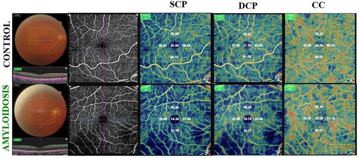
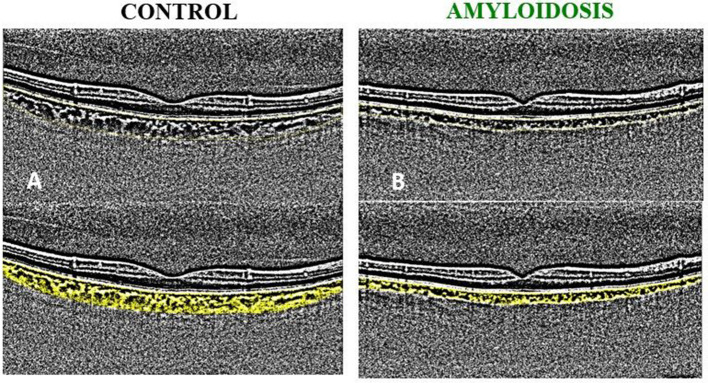
Purpose: Retinal angiopathy represents a well-known ocular manifestation of hereditary transthyretin amyloidosis (ATTRv). Until recently, there have been no reports on retinal changes in ATTRwt. In this retrospective observational clinical study, we aimed to determine whether vessel density (VD) indices and the choroidal vascularity index (CVI) could offer insights into retinal and choroidal vascular changes among patients affected by ATTRwt.
Methods: Eighteen patients with a confirmed diagnosis of ATTRwt underwent structural optical coherence tomography (OCT) and OCT angiography (OCTA). We established a control group consisting of 16 healthy subjects for statistical comparisons. The 3D OCT and OCTA datasets were analyzed to assess retinal and choroidal thickness and VD. For measuring CVI, we obtained measurements for the total choroid area (TCA), luminal area (LA), and stromal area (SA).
Results: The mean VD exhibited a statistically significant reduction in the superficial capillary plexus (SCP), deep capillary plexus (DCP), and choriocapillaris (CC) among the ATTRwt group in comparison to the control group (p < 0.0001). Notably, ATTRwt patients displayed decreased choroidal thickness (p = 0.08). Additionally, the median CVI was lower in the ATTRwt group than in the control group (p = 0.04).
Conclusion: The indices from OCTA and CVI have the potential to serve as non-invasive biomarkers for the quantitative evaluation of retinal and choroidal vascular involvement in patients with ATTRwt.
Keywords: OCT angiography (OCTA); choroidal vascularity index (CVI); retinal and choroidal microangiopathy; retinal vessel density; wild-type (ATTRwt) transthyretin amyloidosis.
Copyright © 2024 Rinaldi, Tranfa, Chiosi, Campagna, De Bernardo, Gioia, Natale, Caiazza, Dongiglio, Verrillo, Palmiero, Limongelli and Costagliola.
Conflict of interest statement
The authors declare that the research was conducted in the absence of any commercial or financial relationships that could be construed as a potential conflict of interest.
Figures
References
LinkOut - more resources
Full Text Sources
Research Materials
Miscellaneous